 Movie
Movie Controller

[English] 日本語
 Yorodumi
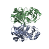
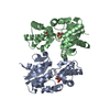
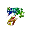
- PDB-1b2p: NATIVE MANNOSE-SPECIFIC BULB LECTIN FROM SCILLA CAMPANULATA (BLUE... -
+ Open data

- Basic information
| Entry | Database: PDB / ID: 1b2p | ||||||
|---|---|---|---|---|---|---|---|
| Title | NATIVE MANNOSE-SPECIFIC BULB LECTIN FROM SCILLA CAMPANULATA (BLUEBELL) AT 1.7 ANGSTROMS RESOLUTION | ||||||
 Components Components | PROTEIN (LECTIN) | ||||||
 Keywords Keywords | SUGAR BINDING PROTEIN / MANNOSE-BINDING LECTIN / MONOCOT / AGLUTININ / BLUEBELL BULBS / PROTEIN- CARBOHYDRATE INTERACTIONS | ||||||
| Function / homology |  Function and homology information Function and homology information | ||||||
| Biological species |  Hyacinthoides hispanica (Spanish bluebell) Hyacinthoides hispanica (Spanish bluebell) | ||||||
| Method |  X-RAY DIFFRACTION / SYNCHROTRON / MOLECULAR REPLACEMENT / Resolution: 1.7 Å X-RAY DIFFRACTION / SYNCHROTRON / MOLECULAR REPLACEMENT / Resolution: 1.7 Å | ||||||
 Authors Authors | Wood, S.D. / Wright, L.M. / Reynolds, C.D. / Rizkallah, P.J. / Allen, A.K. / Peumans, W.J. / Van Damme, E.J.M. | ||||||
 Citation Citation | Journal: Acta Crystallogr.,Sect.D / Year: 1999 Title: Structure of the native (unligated) mannose-specific bulb lectin from Scilla campanulata (bluebell) at 1.7 A resolution. Authors: Wood, S.D. / Wright, L.M. / Reynolds, C.D. / Rizkallah, P.J. / Allen, A.K. / Peumans, W.J. / Van Damme, E.J. #1: Journal: Acta Crystallogr.,Sect.D / Year: 1998Title: Crystallisation and Preliminary Structural Studies of Scilla Campanulata Lectin Complexed with Alpha (1-6)-Mannobiose Authors: Wright, L.M. / Wood, S.D. / Reynolds, C.D. / Rizkallah, P.J. / Allen, A.K. #2: Journal: Acta Crystallogr.,Sect.D / Year: 1998Title: Scilla Campanulata Agglutinin Crystallized in Complex with the Trimannoside Alpha-D-Man-(1-6)-[Alpha-D-Man-(1-3)]-Alpha-D-Man Authors: Wright, L.M. / Rizkallah, P.J. / Wood, S.D. / Reynolds, C.D. #3: Journal: Protein Pept.Lett. / Year: 1997Title: Crystallisation and Preliminary Crystallographic Analysis of Scilla Campanulata Lectin Complexed with Alpha-D-Mannose Authors: Wright, L.M. / Wood, S.D. / Reynolds, C.D. / Rizkallah, P.J. / Allen, A.K. #4: Journal: Acta Crystallogr.,Sect.D / Year: 1996Title: Purification, Crystallisation and Preliminary X-Ray Analysis of a Mannose- Binding Lectin from Bluebell (Scilla Campanulata) Bulbs Authors: Wright, L.M. / Wood, S.D. / Reynolds, C.D. / Rizkallah, P.J. / Peumans, W.J. / Vandamme, E.J.M. / Allen, A.K. | ||||||
| History |
|
- Structure visualization
| Structure viewer | Molecule: MolmilJmol/JSmol |
|---|
- Downloads & links
-Download
| PDBx/mmCIF format | 1b2p.cif.gz | 64.6 KB | Display | PDBx/mmCIF format |
|---|---|---|---|---|
| PDB format | pdb1b2p.ent.gz | 47.7 KB | Display | PDB format |
| PDBx/mmJSON format | 1b2p.json.gz | Tree view | PDBx/mmJSON format | |
| Others |  Other downloads Other downloads |
-Validation report
| Arichive directory | https://data.pdbj.org/pub/pdb/validation_reports/b2/1b2pftp://data.pdbj.org/pub/pdb/validation_reports/b2/1b2p | HTTPS FTP |
|---|
-Related structure data
| Related structure data |  1gnaS S: Starting model for refinement |
|---|---|
| Similar structure data |


-Links
 PDBj
PDBj
- Assembly
| Deposited unit | 
| ||||||||
|---|---|---|---|---|---|---|---|---|---|
| 1 | 
| ||||||||
| 2 |
| ||||||||
| Unit cell |
|
-Components
| #1: Protein | Mass: 13209.855 Da / Num. of mol.: 2 / Source method: isolated from a natural source Details: LOCATED IN THE BULBS OF SCILLA CAMPANULATA (BLUEBELL) Source: (natural) Hyacinthoides hispanica (Spanish bluebell)References: UniProt: Q9ZP49 #2: Water | ChemComp-HOH / |  Mass: 18.015 Da / Num. of mol.: 245 / Source method: isolated from a natural source / Formula: H2O Mass: 18.015 Da / Num. of mol.: 245 / Source method: isolated from a natural source / Formula: H2OHas protein modification | Y | |
|---|
-Experimental details
-Experiment
| Experiment | Method: X-RAY DIFFRACTION / Number of used crystals: 1 |
|---|
- Sample preparation
| Crystal | Density Matthews: 2.78 Å3/Da / Density % sol: 56 % | |||||||||||||||||||||||||
|---|---|---|---|---|---|---|---|---|---|---|---|---|---|---|---|---|---|---|---|---|---|---|---|---|---|---|
| Crystal grow | Method: vapor diffusion, hanging drop / pH: 4.7 Details: HANGING-DROP VAPOUR-DIFFUSION METHOD WELL: 70% SATURATED AMMONIUM SULPHATE, PH 4.7 DROP: 5.5 MG/ML PROTEIN, 10MM DAP, 600MM PHOSPHATE BUFFERED SALINE, VAPOR DIFFUSION, HANGING DROP | |||||||||||||||||||||||||
| Components of the solutions |
| |||||||||||||||||||||||||
| Crystal | *PLUS | |||||||||||||||||||||||||
| Crystal grow | *PLUS | |||||||||||||||||||||||||
| Components of the solutions | *PLUS
|
-Data collection
| Diffraction | Mean temperature: 277 K |
|---|---|
| Diffraction source | Source: SYNCHROTRON / Site: SRS  / Beamline: PX9.5 / Wavelength: 0.8 / Beamline: PX9.5 / Wavelength: 0.8 |
| Detector | Type: MARRESEARCH / Detector: IMAGE PLATE / Date: Feb 13, 1996 / Details: MIRRORS |
| Radiation | Monochromator: NI FILTER / Protocol: SINGLE WAVELENGTH / Monochromatic (M) / Laue (L): M / Scattering type: x-ray |
| Radiation wavelength | Wavelength: 0.8 Å / Relative weight: 1 |
| Reflection | Resolution: 1.7→20 Å / Num. obs: 33837 / % possible obs: 98.5 % / Observed criterion σ(I): 2 / Redundancy: 3.8 % / Rmerge(I) obs: 0.039 / Rsym value: 0.1 |
| Reflection shell | Highest resolution: 1.7 Å |
| Reflection | *PLUS Num. measured all: 161054 |
| Reflection shell | *PLUS Mean I/σ(I) obs: 3.2 |
- Processing
| Software |
| |||||||||||||||||||||||||||||||||||||||||||||||||||||||||||||||
|---|---|---|---|---|---|---|---|---|---|---|---|---|---|---|---|---|---|---|---|---|---|---|---|---|---|---|---|---|---|---|---|---|---|---|---|---|---|---|---|---|---|---|---|---|---|---|---|---|---|---|---|---|---|---|---|---|---|---|---|---|---|---|---|---|
| Refinement | Method to determine structure: MOLECULAR REPLACEMENT Starting model: 1GNA Resolution: 1.7→20 Å / Cross valid method: THROUGHOUT / σ(F): 0 / ESU R: 0.221 / ESU R Free: 0.225 / Details: X-PLOR WAS USED INITIALLY
| |||||||||||||||||||||||||||||||||||||||||||||||||||||||||||||||
| Displacement parameters | Biso mean: 20.5 Å2 | |||||||||||||||||||||||||||||||||||||||||||||||||||||||||||||||
| Refinement step | Cycle: LAST / Resolution: 1.7→20 Å
| |||||||||||||||||||||||||||||||||||||||||||||||||||||||||||||||
| Refine LS restraints |
| |||||||||||||||||||||||||||||||||||||||||||||||||||||||||||||||
| Software | *PLUS Name: REFMAC / Classification: refinement | |||||||||||||||||||||||||||||||||||||||||||||||||||||||||||||||
| Refinement | *PLUS Highest resolution: 1.7 Å / σ(F): 2 / % reflection Rfree: 5 % / Rfactor obs: 0.186 | |||||||||||||||||||||||||||||||||||||||||||||||||||||||||||||||
| Solvent computation | *PLUS | |||||||||||||||||||||||||||||||||||||||||||||||||||||||||||||||
| Displacement parameters | *PLUS Biso mean: 20.5 Å2 | |||||||||||||||||||||||||||||||||||||||||||||||||||||||||||||||
| Refine LS restraints | *PLUS
|
