Evidence: homology, Homologous to the glucose 1,6-bisphospate complex in the D10N variant (PDB: 5ok1). Heteronuclear NMR was performed to confirm the binding interaction in solution.
Type
Name
Symmetry operation
Number
identity operation
1_555
x,y,z
1
Buried area
1270 Å2
ΔGint
-17 kcal/mol
Surface area
20770 Å2
Method
PISA
Unit cell
Length a, b, c (Å)
38.270, 117.330, 52.780
Angle α, β, γ (deg.)
90.00, 97.78, 90.00
Int Tables number
4
Space group name H-M
P1211
-
Components
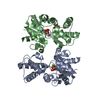
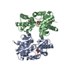
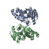
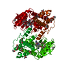
#1: Protein
Beta-phosphoglucomutase / Beta-PGM
Mass: 24239.594 Da / Num. of mol.: 2 Source method: isolated from a genetically manipulated source Source: (gene. exp.) Lactococcus lactis subsp. lactis (strain IL1403) (lactic acid bacteria) Gene: pgmB, LL0429, L0001 / Production host: Escherichia coli BL21(DE3) (bacteria) / References: UniProt: P71447, beta-phosphoglucomutase
Resolution: 2.47→58.66 Å / Cor.coef. Fo:Fc: 0.948 / Cor.coef. Fo:Fc free: 0.867 / SU B: 12.567 / SU ML: 0.269 / Cross valid method: THROUGHOUT / ESU R: 0.785 / ESU R Free: 0.334 / Details: HYDROGENS HAVE BEEN ADDED IN THE RIDING POSITIONS
Rfactor
Num. reflection
% reflection
Selection details
Rfree
0.27827
832
5 %
RANDOM
Rwork
0.19301
-
-
-
obs
0.19723
15698
99.74 %
-
Solvent computation
Ion probe radii: 0.8 Å / Shrinkage radii: 0.8 Å / VDW probe radii: 1.2 Å
 Movie
Movie Controller

 Yorodumi
 Open data
 Basic information
 Components
 Keywords
 Function and homology information
 Lactococcus lactis subsp. lactis (lactic acid bacteria)
 X-RAY DIFFRACTION /
  Authors
 United Kingdom, 2items
  Citation
 Structure visualization
 Downloads & links
 Other downloads
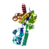


 PDBj
PDBj

 Assembly

 Mass: 24.305 Da / Num. of mol.: 2 / Source method: obtained synthetically / Formula: Mg
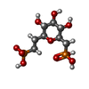
 Type: D-saccharide / Mass: 336.170 Da / Num. of mol.: 2 / Source method: obtained synthetically / Formula: C8H18O10P2
 Mass: 18.015 Da / Num. of mol.: 142 / Source method: isolated from a natural source / Formula: H2O
 Sample preparation
 Processing