 Movie
Movie Controller

[English] 日本語
 Yorodumi
- PDB-1b2m: THREE-DIMENSIONAL STRUCTURE OF RIBONULCEASE T1 COMPLEXED WITH AN ... -
+ Open data

- Basic information
| Entry | Database: PDB / ID: 1b2m | ||||||
|---|---|---|---|---|---|---|---|
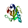
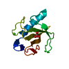
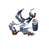
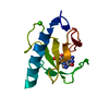
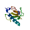
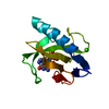
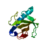
| Title | THREE-DIMENSIONAL STRUCTURE OF RIBONULCEASE T1 COMPLEXED WITH AN ISOSTERIC PHOSPHONATE ANALOGUE OF GPU: ALTERNATE SUBSTRATE BINDING MODES AND CATALYSIS. | ||||||
 Components Components |
| ||||||
 Keywords Keywords | HYDROLASE/RNA / HYDROLASE / ENDORIBONUCLEASE / HYDROLASE-RNA complex | ||||||
| Function / homology |  Function and homology information Function and homology informationhyphal tip / ribonuclease T1 / ribonuclease T1 activity / cell septum / hydrolase activity / RNA binding Similarity search - Function | ||||||
| Biological species |  | ||||||
| Method |  X-RAY DIFFRACTION / MOLECULAR REPLACEMENT / Resolution: 2 Å X-RAY DIFFRACTION / MOLECULAR REPLACEMENT / Resolution: 2 Å | ||||||
 Authors Authors | Arni, R.K. / Watanabe, L. / Ward, R.J. / Kreitman, R.J. / Kumar, K. / Walz Jr., F.G. | ||||||
 Citation Citation | Journal: Biochemistry / Year: 1999 Title: Three-dimensional structure of ribonuclease T1 complexed with an isosteric phosphonate substrate analogue of GpU: alternate substrate binding modes and catalysis. Authors: Arni, R.K. / Watanabe, L. / Ward, R.J. / Kreitman, R.J. / Kumar, K. / Walz Jr., F.G. #1: Journal: Biochemistry / Year: 1992Title: Three-Dimensional Structure of Gln 25-Ribonuclease T1 at 1.84 Angstroms Resolution: Structural Variations at the Base Recognition and Catalytic Sites Authors: Arni, R.K. / Pal, G.P. / Ravichandran, K.G. / Tulinsky, A. / Metcalf Jr., P.F.G.W. #2: Journal: Biochemistry / Year: 1989Title: Crystal Structure of Guanosine-Free Ribonuclease T1, Complexed with Vanadate(V), Suggests Conformational Change Upon Substrate Binding Authors: Kostrewa, D. / Choe, H.-W. / Heinemann, U. / Saenger, W. #3: Journal: J.Biol.Chem. / Year: 1988Title: Three Dimensional Structures of the Ribonuclease T1 2'-Gmp Complex at 1.9 Angstroms Resolution Authors: Arni, R. / Heinemann, U. / Tokuoka, R. / Saenger, W. #4: Journal: Acta Crystallogr.,Sect.B / Year: 1987Title: Restrained Least-Squares Refinement of the Crystal Structure of the Ribonuclease T1 2'-Guanylic Acid Complex at 1.9 Angstroms Resolution Authors: Arni, R. / Heinemann, V. / Maslowska, M. / Tokuoka, R. / Saenger, W. #5: Journal: Fresenius Z.Anal.Chem. / Year: 1987Title: Structure and Function of the Enzyme Ribonuclease T1 Authors: Arni, R. / Heinemann, U. / Saenger, W. #6: Journal: J.Mol.Biol. / Year: 1980Title: Crystallization of Ribonuclease T1 Authors: Martin, P.O. / Tulinsky, A. / Walz, F.G. | ||||||
| History |
|
- Structure visualization
| Structure viewer | Molecule: MolmilJmol/JSmol |
|---|
- Downloads & links
-Download
| PDBx/mmCIF format | 1b2m.cif.gz | 57 KB | Display | PDBx/mmCIF format |
|---|---|---|---|---|
| PDB format | pdb1b2m.ent.gz | 41.1 KB | Display | PDB format |
| PDBx/mmJSON format | 1b2m.json.gz | Tree view | PDBx/mmJSON format | |
| Others |  Other downloads Other downloads |
-Validation report
| Arichive directory | https://data.pdbj.org/pub/pdb/validation_reports/b2/1b2mftp://data.pdbj.org/pub/pdb/validation_reports/b2/1b2m | HTTPS FTP |
|---|
-Related structure data
| Related structure data | 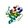 1rntS S: Starting model for refinement |
|---|---|
| Similar structure data |


-Links
 PDBj
PDBj- Assembly
| Deposited unit | 
| ||||||||||||
|---|---|---|---|---|---|---|---|---|---|---|---|---|---|
| 1 | 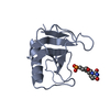
| ||||||||||||
| 2 | 
| ||||||||||||
| Unit cell |
| ||||||||||||
| Noncrystallographic symmetry (NCS) | NCS oper:
|
-Components
| #1: RNA chain | Mass: 620.441 Da / Num. of mol.: 3 / Source method: obtained synthetically #2: Protein | Mass: 11093.644 Da / Num. of mol.: 2 / Mutation: GLN 25 VARIANT / Source method: isolated from a natural source / Details: RNASE T1 COMPLEXED WITH 5'-R(*GP*(CH2)U)-3 / Source: (natural) #3: Water | ChemComp-HOH / |  Mass: 18.015 Da / Num. of mol.: 92 / Source method: isolated from a natural source / Formula: H2O Mass: 18.015 Da / Num. of mol.: 92 / Source method: isolated from a natural source / Formula: H2OHas protein modification | Y | |
|---|
-Experimental details
-Experiment
| Experiment | Method: X-RAY DIFFRACTION / Number of used crystals: 1 |
|---|
- Sample preparation
| Crystal | Density Matthews: 2.23 Å3/Da / Density % sol: 44.82 % | ||||||||||||||||||||||||||||||||||||
|---|---|---|---|---|---|---|---|---|---|---|---|---|---|---|---|---|---|---|---|---|---|---|---|---|---|---|---|---|---|---|---|---|---|---|---|---|---|
| Crystal grow | pH: 4.1 / Details: pH 4.10 | ||||||||||||||||||||||||||||||||||||
| Components of the solutions |
| ||||||||||||||||||||||||||||||||||||
| Crystal grow | *PLUS pH: 4.1 / Method: vapor diffusion, hanging drop | ||||||||||||||||||||||||||||||||||||
| Components of the solutions | *PLUS
|
-Data collection
| Diffraction | Mean temperature: 287 K |
|---|---|
| Diffraction source | Source: ROTATING ANODE / Type: RIGAKU RU200 / Wavelength: 1.5418 |
| Detector | Type: RIGAKU / Detector: IMAGE PLATE |
| Radiation | Monochromator: NI FILTER / Protocol: SINGLE WAVELENGTH / Monochromatic (M) / Laue (L): M / Scattering type: x-ray |
| Radiation wavelength | Wavelength: 1.5418 Å / Relative weight: 1 |
| Reflection | Resolution: 2→20 Å / Num. obs: 11298 / % possible obs: 75 % / Observed criterion σ(I): 2 / Rsym value: 0.64 |
| Reflection | *PLUS Highest resolution: 2 Å / Lowest resolution: 20 Å / % possible obs: 75 % / Observed criterion σ(I): 2 / Rmerge(I) obs: 0.064 |
- Processing
| Software |
| ||||||||||||||||||||||||||||||||||||||||||||||||||||||||||||
|---|---|---|---|---|---|---|---|---|---|---|---|---|---|---|---|---|---|---|---|---|---|---|---|---|---|---|---|---|---|---|---|---|---|---|---|---|---|---|---|---|---|---|---|---|---|---|---|---|---|---|---|---|---|---|---|---|---|---|---|---|---|
| Refinement | Method to determine structure: MOLECULAR REPLACEMENT Starting model: 1RNT Resolution: 2→8 Å / σ(F): 2
| ||||||||||||||||||||||||||||||||||||||||||||||||||||||||||||
| Refinement step | Cycle: LAST / Resolution: 2→8 Å
| ||||||||||||||||||||||||||||||||||||||||||||||||||||||||||||
| Refine LS restraints |
| ||||||||||||||||||||||||||||||||||||||||||||||||||||||||||||
| Software | *PLUS Name: X-PLOR / Version: 3.1 / Classification: refinement | ||||||||||||||||||||||||||||||||||||||||||||||||||||||||||||
| Refinement | *PLUS Highest resolution: 2 Å / Lowest resolution: 8 Å / Num. reflection obs: 26734 | ||||||||||||||||||||||||||||||||||||||||||||||||||||||||||||
| Solvent computation | *PLUS | ||||||||||||||||||||||||||||||||||||||||||||||||||||||||||||
| Displacement parameters | *PLUS | ||||||||||||||||||||||||||||||||||||||||||||||||||||||||||||
| Refine LS restraints | *PLUS
|
