 Movie
Movie Controller


+ Open data

- Basic information
| Entry | Database: PDB / ID: 1bu4 | ||||||
|---|---|---|---|---|---|---|---|
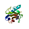
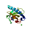
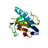
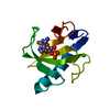
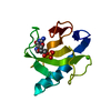
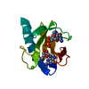
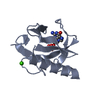
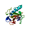
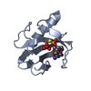
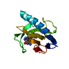
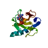
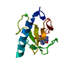
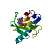
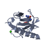
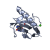
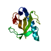
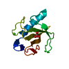
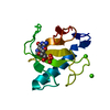
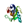
| Title | RIBONUCLEASE 1 COMPLEX WITH 2'GMP | ||||||
 Components Components | RIBONUCLEASE T1 | ||||||
 Keywords Keywords | ENDORIBONUCLEASE / HYDROLASE | ||||||
| Function / homology |  Function and homology information Function and homology informationhyphal tip / ribonuclease T1 / ribonuclease T1 activity / cell septum / hydrolase activity / RNA binding Similarity search - Function | ||||||
| Biological species |  | ||||||
| Method |  X-RAY DIFFRACTION / MOLECULAR REPLACEMENT / Resolution: 1.9 Å X-RAY DIFFRACTION / MOLECULAR REPLACEMENT / Resolution: 1.9 Å | ||||||
 Authors Authors | Loris, R. / Devos, S. / Langhorst, U. / Decanniere, K. / Bouckaert, J. / Maes, D. / Transue, T.R. / Steyaert, J. | ||||||
 Citation Citation | Journal: Proteins / Year: 1999 Title: Conserved water molecules in a large family of microbial ribonucleases. Authors: Loris, R. / Langhorst, U. / De Vos, S. / Decanniere, K. / Bouckaert, J. / Maes, D. / Transue, T.R. / Steyaert, J. | ||||||
| History |
|
- Structure visualization
| Structure viewer | Molecule: MolmilJmol/JSmol |
|---|
- Downloads & links
-Download
| PDBx/mmCIF format | 1bu4.cif.gz | 34.7 KB | Display | PDBx/mmCIF format |
|---|---|---|---|---|
| PDB format | pdb1bu4.ent.gz | 22.1 KB | Display | PDB format |
| PDBx/mmJSON format | 1bu4.json.gz | Tree view | PDBx/mmJSON format | |
| Others |  Other downloads Other downloads |
-Validation report
| Arichive directory | https://data.pdbj.org/pub/pdb/validation_reports/bu/1bu4ftp://data.pdbj.org/pub/pdb/validation_reports/bu/1bu4 | HTTPS FTP |
|---|
-Related structure data


-Links
 PDBj
PDBj- Assembly
| Deposited unit | 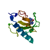
| ||||||||
|---|---|---|---|---|---|---|---|---|---|
| 1 |
| ||||||||
| Unit cell |
|
-Components
| #1: Protein | Mass: 11094.694 Da / Num. of mol.: 1 Source method: isolated from a genetically manipulated source Source: (gene. exp.)  |
|---|---|
| #2: Chemical | ChemComp-CA /   Mass: 40.078 Da / Num. of mol.: 1 / Source method: obtained synthetically / Formula: Ca Mass: 40.078 Da / Num. of mol.: 1 / Source method: obtained synthetically / Formula: Ca |
| #3: Chemical | ChemComp-2GP /   Mass: 363.221 Da / Num. of mol.: 1 / Source method: obtained synthetically / Formula: C10H14N5O8P Mass: 363.221 Da / Num. of mol.: 1 / Source method: obtained synthetically / Formula: C10H14N5O8P |
| #4: Water | ChemComp-HOH /  Mass: 18.015 Da / Num. of mol.: 79 / Source method: isolated from a natural source / Formula: H2O Mass: 18.015 Da / Num. of mol.: 79 / Source method: isolated from a natural source / Formula: H2O |
| Has protein modification | Y |
-Experimental details
-Experiment
| Experiment | Method: X-RAY DIFFRACTION / Number of used crystals: 1 |
|---|
- Sample preparation
| Crystal | Density Matthews: 2.26 Å3/Da / Density % sol: 46 % | ||||||||||||||||||||||||||||||||||||
|---|---|---|---|---|---|---|---|---|---|---|---|---|---|---|---|---|---|---|---|---|---|---|---|---|---|---|---|---|---|---|---|---|---|---|---|---|---|
| Crystal grow | pH: 4.5 / Details: pH 4.5 | ||||||||||||||||||||||||||||||||||||
| Crystal grow | *PLUS pH: 4.2 / Method: vapor diffusion, hanging drop / Details: Zegers, I., (1998) Nat. Struct. Biol., 5, 280. | ||||||||||||||||||||||||||||||||||||
| Components of the solutions | *PLUS
|
-Data collection
| Diffraction | Mean temperature: 293 K |
|---|---|
| Diffraction source | Source: ROTATING ANODE / Type: RIGAKU RUH2R / Wavelength: 1.5418 |
| Detector | Type: MARRESEARCH / Detector: IMAGE PLATE / Date: Feb 1, 1998 |
| Radiation | Monochromator: GRAPHITE(002) / Monochromatic (M) / Laue (L): M / Scattering type: x-ray |
| Radiation wavelength | Wavelength: 1.5418 Å / Relative weight: 1 |
| Reflection | Resolution: 1.9→20 Å / Num. obs: 7907 / % possible obs: 95.6 % / Redundancy: 5.5 % / Rsym value: 0.139 / Net I/σ(I): 12.66 |
| Reflection shell | Resolution: 1.9→1.95 Å / Redundancy: 5.5 % / Mean I/σ(I) obs: 2.47 / Rsym value: 0.583 / % possible all: 92.2 |
| Reflection | *PLUS Num. obs: 7853 / Redundancy: 5.6 % / Num. measured all: 44006 / Rmerge(I) obs: 0.139 |
| Reflection shell | *PLUS Rmerge(I) obs: 0.583 |
- Processing
| Software |
| ||||||||||||||||||||
|---|---|---|---|---|---|---|---|---|---|---|---|---|---|---|---|---|---|---|---|---|---|
| Refinement | Method to determine structure: MOLECULAR REPLACEMENT Starting model: PDB ENTRY Resolution: 1.9→20 Å / Cross valid method: THROUGHOUT / σ(F): 0
| ||||||||||||||||||||
| Refinement step | Cycle: LAST / Resolution: 1.9→20 Å
| ||||||||||||||||||||
| Software | *PLUS Name: REFMAC / Classification: refinement | ||||||||||||||||||||
| Refinement | *PLUS Rfactor obs: 0.18 / Rfactor Rfree: 0.23 | ||||||||||||||||||||
| Solvent computation | *PLUS | ||||||||||||||||||||
| Displacement parameters | *PLUS | ||||||||||||||||||||
| Refine LS restraints | *PLUS
|
