 Movie
Movie Controller


+ Open data

- Basic information
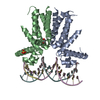
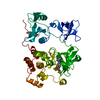
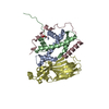
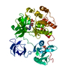
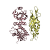
| Entry | Database: PDB / ID: 1awc | ||||||
|---|---|---|---|---|---|---|---|
| Title | MOUSE GABP ALPHA/BETA DOMAIN BOUND TO DNA | ||||||
 Components Components |
| ||||||
 Keywords Keywords | TRANSCRIPTION/DNA / COMPLEX (TRANSCRIPTION REGULATION-DNA) / DNA-BINDING / NUCLEAR PROTEIN / ETS DOMAIN / ANKYRIN REPEATS / TRANSCRIPTION FACTOR / TRANSCRIPTION-DNA COMPLEX | ||||||
| Function / homology |  Function and homology information Function and homology informationTranscriptional activation of mitochondrial biogenesis / blastocyst formation / ERBB signaling pathway / neuromuscular junction development / negative regulation of megakaryocyte differentiation / mitochondrion organization / in utero embryonic development / cytoplasmic ribonucleoprotein granule / sequence-specific double-stranded DNA binding / DNA-binding transcription activator activity, RNA polymerase II-specific ...Transcriptional activation of mitochondrial biogenesis / blastocyst formation / ERBB signaling pathway / neuromuscular junction development / negative regulation of megakaryocyte differentiation / mitochondrion organization / in utero embryonic development / cytoplasmic ribonucleoprotein granule / sequence-specific double-stranded DNA binding / DNA-binding transcription activator activity, RNA polymerase II-specific / DNA-binding transcription factor activity, RNA polymerase II-specific / cell differentiation / transcription cis-regulatory region binding / RNA polymerase II cis-regulatory region sequence-specific DNA binding / chromatin binding / regulation of transcription by RNA polymerase II / chromatin / negative regulation of transcription by RNA polymerase II / positive regulation of transcription by RNA polymerase II / DNA-templated transcription / nucleoplasm / nucleus Similarity search - Function | ||||||
| Biological species |  | ||||||
| Method |  X-RAY DIFFRACTION / SYNCHROTRON / MIR / Resolution: 2.15 Å X-RAY DIFFRACTION / SYNCHROTRON / MIR / Resolution: 2.15 Å | ||||||
 Authors Authors | Batchelor, A.H. / Wolberger, C. | ||||||
 Citation Citation | Journal: Science / Year: 1998 Title: The structure of GABPalpha/beta: an ETS domain- ankyrin repeat heterodimer bound to DNA. Authors: Batchelor, A.H. / Piper, D.E. / de la Brousse, F.C. / McKnight, S.L. / Wolberger, C. | ||||||
| History |
|
- Structure visualization
| Structure viewer | Molecule: MolmilJmol/JSmol |
|---|
- Downloads & links
-Download
| PDBx/mmCIF format | 1awc.cif.gz | 89.8 KB | Display | PDBx/mmCIF format |
|---|---|---|---|---|
| PDB format | pdb1awc.ent.gz | 65.1 KB | Display | PDB format |
| PDBx/mmJSON format | 1awc.json.gz | Tree view | PDBx/mmJSON format | |
| Others |  Other downloads Other downloads |
-Validation report
| Arichive directory | https://data.pdbj.org/pub/pdb/validation_reports/aw/1awcftp://data.pdbj.org/pub/pdb/validation_reports/aw/1awc | HTTPS FTP |
|---|
-Related structure data
| Similar structure data |
|---|


-Links
 PDBj
PDBj


- Assembly
| Deposited unit | 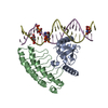
| ||||||||||
|---|---|---|---|---|---|---|---|---|---|---|---|
| 1 |
| ||||||||||
| Unit cell |
|
-Components
| #1: DNA chain | Mass: 6712.888 Da / Num. of mol.: 1 / Source method: obtained synthetically |
|---|---|
| #2: DNA chain | Mass: 6525.857 Da / Num. of mol.: 1 / Source method: obtained synthetically |
| #3: Protein | Mass: 13065.230 Da / Num. of mol.: 1 / Fragment: ETS DOMAIN PLUS 30 C-TERMINAL RESIDUES Source method: isolated from a genetically manipulated source Source: (gene. exp.)  |
| #4: Protein | Mass: 16834.010 Da / Num. of mol.: 1 / Fragment: ANKYRIN REPEAT DOMAIN Source method: isolated from a genetically manipulated source Source: (gene. exp.) |
| #5: Water | ChemComp-HOH /  Mass: 18.015 Da / Num. of mol.: 46 / Source method: isolated from a natural source / Formula: H2O Mass: 18.015 Da / Num. of mol.: 46 / Source method: isolated from a natural source / Formula: H2O |
-Experimental details
-Experiment
| Experiment | Method: X-RAY DIFFRACTION / Number of used crystals: 1 |
|---|
- Sample preparation
| Crystal | Density Matthews: 2.4 Å3/Da / Density % sol: 49 % | ||||||||||||||||||||||||||||||||||||||||||||||||||||||||||||
|---|---|---|---|---|---|---|---|---|---|---|---|---|---|---|---|---|---|---|---|---|---|---|---|---|---|---|---|---|---|---|---|---|---|---|---|---|---|---|---|---|---|---|---|---|---|---|---|---|---|---|---|---|---|---|---|---|---|---|---|---|---|
| Crystal grow | pH: 9 Details: 100 MM BIS-TRIS PROPANE PH 9, 12 % PEG 1000, 5 MM COBALTIC HEXAMINE CHLORIDE, 1 MM DTT, 20 MM TRIS, 1 MM EDTA, 0.001 % SODIUM AZIDE, pH 9.0 | ||||||||||||||||||||||||||||||||||||||||||||||||||||||||||||
| Components of the solutions |
| ||||||||||||||||||||||||||||||||||||||||||||||||||||||||||||
| Crystal | *PLUS Density % sol: 49 % | ||||||||||||||||||||||||||||||||||||||||||||||||||||||||||||
| Crystal grow | *PLUS Temperature: 20 ℃ / Method: vapor diffusion, hanging drop | ||||||||||||||||||||||||||||||||||||||||||||||||||||||||||||
| Components of the solutions | *PLUS
|
-Data collection
| Diffraction | Mean temperature: 100 K |
|---|---|
| Diffraction source | Source: SYNCHROTRON / Site: NSLS  / Beamline: X4A / Beamline: X4A |
| Detector | Type: FUJI / Detector: IMAGE PLATE / Date: May 15, 1996 / Details: SPHERICAL RH COATED |
| Radiation | Monochromator: SAGITALLY FOCUSED SI(111) / Protocol: SINGLE WAVELENGTH / Monochromatic (M) / Laue (L): M / Scattering type: x-ray |
| Radiation wavelength | Relative weight: 1 |
| Reflection | Resolution: 2.15→30 Å / Num. obs: 18368 / % possible obs: 88 % / Observed criterion σ(I): 1.5 / Redundancy: 8.7 % / Rsym value: 0.072 / Net I/σ(I): 21 |
| Reflection shell | Resolution: 2.15→2.23 Å / Redundancy: 7.9 % / Mean I/σ(I) obs: 5.3 / Rsym value: 0.22 / % possible all: 88 |
| Reflection | *PLUS Highest resolution: 2.15 Å / Lowest resolution: 30 Å / % possible obs: 88 % / Redundancy: 8.7 % / Rmerge(I) obs: 0.072 |
| Reflection shell | *PLUS % possible obs: 88 % / Rmerge(I) obs: 0.22 / Mean I/σ(I) obs: 5.3 |
- Processing
| Software |
| ||||||||||||||||||||||||||||||||||||||||||||||||||||||||||||
|---|---|---|---|---|---|---|---|---|---|---|---|---|---|---|---|---|---|---|---|---|---|---|---|---|---|---|---|---|---|---|---|---|---|---|---|---|---|---|---|---|---|---|---|---|---|---|---|---|---|---|---|---|---|---|---|---|---|---|---|---|---|
| Refinement | Method to determine structure: MIR / Resolution: 2.15→6 Å / Rfactor Rfree error: 0.7 / Data cutoff high absF: 100000 / Data cutoff low absF: 0.1 / Isotropic thermal model: RESTRAINED / Cross valid method: THROUGHOUT
| ||||||||||||||||||||||||||||||||||||||||||||||||||||||||||||
| Displacement parameters | Biso mean: 35.3 Å2
| ||||||||||||||||||||||||||||||||||||||||||||||||||||||||||||
| Refine analyze |
| ||||||||||||||||||||||||||||||||||||||||||||||||||||||||||||
| Refinement step | Cycle: LAST / Resolution: 2.15→6 Å
| ||||||||||||||||||||||||||||||||||||||||||||||||||||||||||||
| Refine LS restraints |
| ||||||||||||||||||||||||||||||||||||||||||||||||||||||||||||
| LS refinement shell | Resolution: 2.15→2.22 Å / Rfactor Rfree error: 0.02 / Total num. of bins used: 10
| ||||||||||||||||||||||||||||||||||||||||||||||||||||||||||||
| Xplor file |
| ||||||||||||||||||||||||||||||||||||||||||||||||||||||||||||
| Software | *PLUS Name: X-PLOR / Version: 3.8 / Classification: refinement | ||||||||||||||||||||||||||||||||||||||||||||||||||||||||||||
| Refinement | *PLUS Highest resolution: 2.15 Å / Lowest resolution: 6 Å / σ(F): 2 / % reflection Rfree: 10 % / Rfactor all: 0.222 / Rfactor obs: 0.211 | ||||||||||||||||||||||||||||||||||||||||||||||||||||||||||||
| Solvent computation | *PLUS | ||||||||||||||||||||||||||||||||||||||||||||||||||||||||||||
| Displacement parameters | *PLUS Biso mean: 35.3 Å2 | ||||||||||||||||||||||||||||||||||||||||||||||||||||||||||||
| Refine LS restraints | *PLUS
| ||||||||||||||||||||||||||||||||||||||||||||||||||||||||||||
| LS refinement shell | *PLUS % reflection Rfree: 10 % |
