 Movie
Movie Controller


+ Open data

- Basic information
| Entry | Database: PDB / ID: 1avf | ||||||
|---|---|---|---|---|---|---|---|
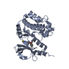
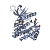
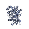
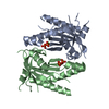
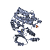
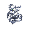
| Title | ACTIVATION INTERMEDIATE 2 OF HUMAN GASTRICSIN FROM HUMAN STOMACH | ||||||
 Components Components | (GASTRICSIN) x 2 | ||||||
 Keywords Keywords | ASPARTYL PROTEASE / GASTRICSIN / ASPARTIC PROTEINASE / INTERMEDIATE / ACTIVATION / ACID | ||||||
| Function / homology |  Function and homology information Function and homology informationgastricsin / positive regulation of antibacterial peptide production / digestion / aspartic-type endopeptidase activity / proteolysis / : Similarity search - Function | ||||||
| Biological species |  Homo sapiens (human) Homo sapiens (human) | ||||||
| Method |  X-RAY DIFFRACTION / SYNCHROTRON / MOLECULAR REPLACEMENT / Resolution: 2.36 Å X-RAY DIFFRACTION / SYNCHROTRON / MOLECULAR REPLACEMENT / Resolution: 2.36 Å | ||||||
 Authors Authors | Khan, A.R. / Cherney, M.M. / Tarasova, N.I. / James, M.N.G. | ||||||
 Citation Citation | Journal: Nat.Struct.Biol. / Year: 1997 Title: Structural characterization of activation 'intermediate 2' on the pathway to human gastricsin. Authors: Khan, A.R. / Cherney, M.M. / Tarasova, N.I. / James, M.N. | ||||||
| History |
|
- Structure visualization
| Structure viewer | Molecule: MolmilJmol/JSmol |
|---|
- Downloads & links
-Download
| PDBx/mmCIF format | 1avf.cif.gz | 151.9 KB | Display | PDBx/mmCIF format |
|---|---|---|---|---|
| PDB format | pdb1avf.ent.gz | 119 KB | Display | PDB format |
| PDBx/mmJSON format | 1avf.json.gz | Tree view | PDBx/mmJSON format | |
| Others |  Other downloads Other downloads |
-Validation report
| Arichive directory | https://data.pdbj.org/pub/pdb/validation_reports/av/1avfftp://data.pdbj.org/pub/pdb/validation_reports/av/1avf | HTTPS FTP |
|---|
-Related structure data
| Related structure data |  1htrS S: Starting model for refinement |
|---|---|
| Similar structure data |


-Links
 PDBj
PDBj
- Assembly
| Deposited unit | 
| ||||||||
|---|---|---|---|---|---|---|---|---|---|
| 1 | 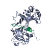
| ||||||||
| 2 | 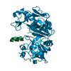
| ||||||||
| Unit cell |
| ||||||||
| Noncrystallographic symmetry (NCS) | NCS oper: (Code: given Matrix: (0.19942, 0.01342, -0.97982), Vector: |
-Components
| #1: Protein/peptide | Mass: 2983.674 Da / Num. of mol.: 2 / Source method: isolated from a natural source / Details: ASPARTIC PROTEINASE ACTIVATION INTERMEDIATE / Source: (natural) Homo sapiens (human) / Organ: STOMACH / Tissue: GASTRIC MUCOSA / References: UniProt: P20142, gastricsin#2: Protein | Mass: 35464.930 Da / Num. of mol.: 2 / Source method: isolated from a natural source / Details: ASPARTIC PROTEINASE ACTIVATION INTERMEDIATE / Source: (natural) Homo sapiens (human) / Organ: STOMACH / Tissue: GASTRIC MUCOSA / References: UniProt: P20142, gastricsin#3: Chemical | ChemComp-NA / |   Mass: 22.990 Da / Num. of mol.: 1 / Source method: obtained synthetically / Formula: Na Mass: 22.990 Da / Num. of mol.: 1 / Source method: obtained synthetically / Formula: Na#4: Water | ChemComp-HOH / |  Mass: 18.015 Da / Num. of mol.: 467 / Source method: isolated from a natural source / Formula: H2O Mass: 18.015 Da / Num. of mol.: 467 / Source method: isolated from a natural source / Formula: H2OHas protein modification | Y | |
|---|
-Experimental details
-Experiment
| Experiment | Method: X-RAY DIFFRACTION / Number of used crystals: 1 |
|---|
- Sample preparation
| Crystal | Density Matthews: 2.5 Å3/Da / Density % sol: 50 % | |||||||||||||||
|---|---|---|---|---|---|---|---|---|---|---|---|---|---|---|---|---|
| Crystal grow | pH: 7.8 Details: PROTEIN WAS CRYSTALLIZED IN 4M NA FORMATE, 100 MM BIS-TRIS-PROPANE, PH 7.8. | |||||||||||||||
| Crystal grow | *PLUS Method: vapor diffusion, hanging drop | |||||||||||||||
| Components of the solutions | *PLUS
|
-Data collection
| Diffraction | Mean temperature: 100 K |
|---|---|
| Diffraction source | Source: SYNCHROTRON / Site: EMBL/DESY, HAMBURG  / Type: EMBL/DESY, HAMBURG / Wavelength: 1 / Type: EMBL/DESY, HAMBURG / Wavelength: 1 |
| Detector | Type: MARRESEARCH / Detector: IMAGE PLATE / Date: Sep 1, 1996 / Details: MIRROR |
| Radiation | Monochromator: GOBEL MIRRORS / Monochromatic (M) / Laue (L): M / Scattering type: x-ray |
| Radiation wavelength | Wavelength: 1 Å / Relative weight: 1 |
| Reflection | Resolution: 2.36→100 Å / Num. obs: 34523 / % possible obs: 95.5 % / Observed criterion σ(I): 2 / Redundancy: 4.3 % / Rmerge(I) obs: 0.066 / Rsym value: 0.117 / Net I/σ(I): 9.2 |
| Reflection shell | Resolution: 2.36→2.4 Å / Redundancy: 2.5 % / Rmerge(I) obs: 0.045 / Mean I/σ(I) obs: 3 / Rsym value: 0.303 / % possible all: 80.8 |
| Reflection shell | *PLUS Lowest resolution: 2.4 Å / % possible obs: 80.8 % |
- Processing
| Software |
| ||||||||||||||||||||||||||||||||||||||||||||||||||||||||||||||||||||||||||||||||
|---|---|---|---|---|---|---|---|---|---|---|---|---|---|---|---|---|---|---|---|---|---|---|---|---|---|---|---|---|---|---|---|---|---|---|---|---|---|---|---|---|---|---|---|---|---|---|---|---|---|---|---|---|---|---|---|---|---|---|---|---|---|---|---|---|---|---|---|---|---|---|---|---|---|---|---|---|---|---|---|---|---|
| Refinement | Method to determine structure: MOLECULAR REPLACEMENT Starting model: PDB ENTRY 1HTR Resolution: 2.36→20 Å / Cross valid method: R-FREE / σ(F): 2
| ||||||||||||||||||||||||||||||||||||||||||||||||||||||||||||||||||||||||||||||||
| Displacement parameters | Biso mean: 18.54 Å2 | ||||||||||||||||||||||||||||||||||||||||||||||||||||||||||||||||||||||||||||||||
| Refine analyze | Luzzati coordinate error obs: 0.3 Å | ||||||||||||||||||||||||||||||||||||||||||||||||||||||||||||||||||||||||||||||||
| Refinement step | Cycle: LAST / Resolution: 2.36→20 Å
| ||||||||||||||||||||||||||||||||||||||||||||||||||||||||||||||||||||||||||||||||
| Refine LS restraints |
| ||||||||||||||||||||||||||||||||||||||||||||||||||||||||||||||||||||||||||||||||
| Refine LS restraints NCS | Weight Biso : 1 / Weight position: 10 | ||||||||||||||||||||||||||||||||||||||||||||||||||||||||||||||||||||||||||||||||
| LS refinement shell | Resolution: 2.36→2.39 Å
| ||||||||||||||||||||||||||||||||||||||||||||||||||||||||||||||||||||||||||||||||
| Software | *PLUS Name: X-PLOR / Classification: refinement | ||||||||||||||||||||||||||||||||||||||||||||||||||||||||||||||||||||||||||||||||
| Refinement | *PLUS Num. reflection all: 34523 / Num. reflection obs: 34124 | ||||||||||||||||||||||||||||||||||||||||||||||||||||||||||||||||||||||||||||||||
| Solvent computation | *PLUS | ||||||||||||||||||||||||||||||||||||||||||||||||||||||||||||||||||||||||||||||||
| Displacement parameters | *PLUS | ||||||||||||||||||||||||||||||||||||||||||||||||||||||||||||||||||||||||||||||||
| Refine LS restraints | *PLUS
|
