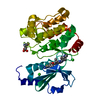
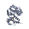
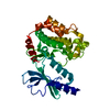
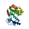
Entry Database : PDB / ID : 3fy0Title Crystal structure of PAK1 kinase domain with ruthenium complex DW1 Serine/threonine-protein kinase PAK 1 Keywords / / / / / / / / / Function / homology Function Domain/homology Component
/ / / / / / / / / / / / / / / / / / / / / / / / / / / / / / / / / / / / / / / / / / / / / / / / / / / / / / / / / / / / / / / / / / / / / / / / / / / / / / / / / / / / / / / / / / / / / / / / / / / / / / / / / / / / / / / / / / / / / / / / / / / / / / / / / Biological species Homo sapiens (human)Method / / / Resolution : 2.35 Å Authors Maksimoska, J. / Marmorstein, R. / Meggers, E. Journal : J.Am.Chem.Soc. / Year : 2008Title : Targeting Large Kinase Active Site with Rigid, Bulky Octahedral Ruthenium ComplexesAuthors : Maksimoska, J. / Feng, L. / Harms, K. / Yi, C. / Kissil, J. / Marmorstein, R. / Meggers, E. History Deposition Jan 21, 2009 Deposition site / Processing site Revision 1.0 Mar 3, 2009 Provider / Type Revision 1.1 Jul 13, 2011 Group / Version format complianceRevision 1.2 Nov 1, 2017 Group / Category / Item Revision 1.3 Oct 20, 2021 Group / Derived calculationsCategory database_2 / struct_conn ... database_2 / struct_conn / struct_ref_seq_dif / struct_site Item _database_2.pdbx_DOI / _database_2.pdbx_database_accession ... _database_2.pdbx_DOI / _database_2.pdbx_database_accession / _struct_conn.pdbx_leaving_atom_flag / _struct_ref_seq_dif.details / _struct_site.pdbx_auth_asym_id / _struct_site.pdbx_auth_comp_id / _struct_site.pdbx_auth_seq_id Revision 1.4 Nov 6, 2024 Group / Structure summaryCategory chem_comp_atom / chem_comp_bond ... chem_comp_atom / chem_comp_bond / pdbx_entry_details / pdbx_modification_feature
Show all Show less
 Movie
Movie Controller

 Yorodumi
 Open data
 Basic information
 Components
 Keywords
 Function and homology information
 Homo sapiens (human)
 X-RAY DIFFRACTION /
  Authors
 Citation
 Structure visualization
 Downloads & links
 Other downloads
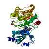


 PDBj
PDBj


 Assembly

 Mass: 496.437 Da / Num. of mol.: 1 / Source method: obtained synthetically / Formula: C23H13N3O4Ru
 Mass: 18.015 Da / Num. of mol.: 119 / Source method: isolated from a natural source / Formula: H2O
 Sample preparation
 / Beamline: 23-ID-B
 Processing