 Movie
Movie Controller


+ Open data

- Basic information
| Entry | Database: PDB / ID: 1akm | ||||||
|---|---|---|---|---|---|---|---|
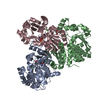
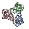
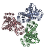
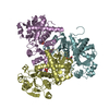
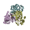
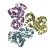
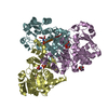
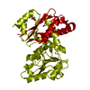
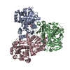
| Title | ORNITHINE TRANSCARBAMYLASE FROM ESCHERICHIA COLI | ||||||
 Components Components | ORNITHINE TRANSCARBAMYLASE | ||||||
 Keywords Keywords | TRANSFERASE / ANABOLIC / UREA CYCLE / CARBAMYL PHOSPHATE | ||||||
| Function / homology |  Function and homology information Function and homology informationornithine carbamoyltransferase / ornithine carbamoyltransferase activity / citrulline biosynthetic process / L-arginine biosynthetic process via ornithine / L-arginine biosynthetic process / amino acid binding / metal ion binding / cytoplasm Similarity search - Function | ||||||
| Biological species |  | ||||||
| Method |  X-RAY DIFFRACTION / MOLECULAR REPLACEMENT, SIRAS / Resolution: 2.8 Å X-RAY DIFFRACTION / MOLECULAR REPLACEMENT, SIRAS / Resolution: 2.8 Å | ||||||
 Authors Authors | Head, J.F. / Seaton, B. / Jin, L. | ||||||
 Citation Citation | Journal: Nat.Struct.Biol. / Year: 1997 Title: Crystal structure at 2.8 A resolution of anabolic ornithine transcarbamylase from Escherichia coli. Authors: Jin, L. / Seaton, B.A. / Head, J.F. | ||||||
| History |
|
- Structure visualization
| Structure viewer | Molecule: MolmilJmol/JSmol |
|---|
- Downloads & links
-Download
| PDBx/mmCIF format | 1akm.cif.gz | 173.7 KB | Display | PDBx/mmCIF format |
|---|---|---|---|---|
| PDB format | pdb1akm.ent.gz | 142.5 KB | Display | PDB format |
| PDBx/mmJSON format | 1akm.json.gz | Tree view | PDBx/mmJSON format | |
| Others |  Other downloads Other downloads |
-Validation report
| Summary document | 1akm_validation.pdf.gz | 440.1 KB | Display | wwPDB validaton report |
|---|---|---|---|---|
| Full document | 1akm_full_validation.pdf.gz | 481.7 KB | Display | |
| Data in XML | 1akm_validation.xml.gz | 38.2 KB | Display | |
| Data in CIF | 1akm_validation.cif.gz | 51 KB | Display | |
| Arichive directory | https://data.pdbj.org/pub/pdb/validation_reports/ak/1akmftp://data.pdbj.org/pub/pdb/validation_reports/ak/1akm | HTTPS FTP |
-Related structure data
| Related structure data | 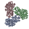 2at2S S: Starting model for refinement |
|---|---|
| Similar structure data |

-Links
 PDBj
PDBj
- Assembly
| Deposited unit | 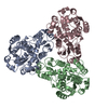
| ||||||||||||
|---|---|---|---|---|---|---|---|---|---|---|---|---|---|
| 1 |
| ||||||||||||
| Unit cell |
| ||||||||||||
| Noncrystallographic symmetry (NCS) | NCS oper:
|
-Components
| #1: Protein | Mass: 36818.840 Da / Num. of mol.: 3 Source method: isolated from a genetically manipulated source Source: (gene. exp.) |
|---|
-Experimental details
-Experiment
| Experiment | Method: X-RAY DIFFRACTION / Number of used crystals: 1 |
|---|
- Sample preparation
| Crystal | Density Matthews: 2.5 Å3/Da / Density % sol: 52 % | ||||||||||||||||||||||||||||||||||||
|---|---|---|---|---|---|---|---|---|---|---|---|---|---|---|---|---|---|---|---|---|---|---|---|---|---|---|---|---|---|---|---|---|---|---|---|---|---|
| Crystal grow | pH: 7.5 Details: CRYSTALLIZED FROM 20% PEG 4000, 10% MPD, 1MM MGSO4, 50MM HEPES, PH 7.5. | ||||||||||||||||||||||||||||||||||||
| Crystal grow | *PLUS Temperature: 17 ℃ / Method: microdialysis | ||||||||||||||||||||||||||||||||||||
| Components of the solutions | *PLUS
|
-Data collection
| Diffraction | Mean temperature: 100 K |
|---|---|
| Diffraction source | Source: ROTATING ANODE / Type: RIGAKU RUH3R / Wavelength: 1.5418 |
| Detector | Type: RIGAKU RAXIS II / Detector: IMAGE PLATE / Date: Aug 1, 1995 / Details: MIRRORS |
| Radiation | Monochromator: NI FILTER / Monochromatic (M) / Laue (L): M / Scattering type: x-ray |
| Radiation wavelength | Wavelength: 1.5418 Å / Relative weight: 1 |
| Reflection | Resolution: 2.8→90 Å / Num. obs: 25858 / % possible obs: 98.6 % / Redundancy: 6.4 % / Rmerge(I) obs: 0.126 / Net I/σ(I): 7.7 |
| Reflection shell | Resolution: 2.8→2.9 Å / Rmerge(I) obs: 0.3 / Mean I/σ(I) obs: 3.4 / % possible all: 99.9 |
| Reflection shell | *PLUS % possible obs: 99.9 % |
- Processing
| Software |
| ||||||||||||||||||||||||||||||||||||||||||||||||||||||||||||
|---|---|---|---|---|---|---|---|---|---|---|---|---|---|---|---|---|---|---|---|---|---|---|---|---|---|---|---|---|---|---|---|---|---|---|---|---|---|---|---|---|---|---|---|---|---|---|---|---|---|---|---|---|---|---|---|---|---|---|---|---|---|
| Refinement | Method to determine structure: MOLECULAR REPLACEMENT, SIRAS Starting model: PDB ENTRY 2AT2 Resolution: 2.8→6 Å / Data cutoff low absF: 0 / Cross valid method: THROUGHOUT / σ(F): 2
| ||||||||||||||||||||||||||||||||||||||||||||||||||||||||||||
| Refinement step | Cycle: LAST / Resolution: 2.8→6 Å
| ||||||||||||||||||||||||||||||||||||||||||||||||||||||||||||
| Refine LS restraints |
| ||||||||||||||||||||||||||||||||||||||||||||||||||||||||||||
| Xplor file |
| ||||||||||||||||||||||||||||||||||||||||||||||||||||||||||||
| Software | *PLUS Name: X-PLOR / Version: 3.1 / Classification: refinement | ||||||||||||||||||||||||||||||||||||||||||||||||||||||||||||
| Refinement | *PLUS Rfactor all: 0.241 | ||||||||||||||||||||||||||||||||||||||||||||||||||||||||||||
| Solvent computation | *PLUS | ||||||||||||||||||||||||||||||||||||||||||||||||||||||||||||
| Displacement parameters | *PLUS |
