 Movie
Movie Controller

[English] 日本語
 Yorodumi
- PDB-5g1p: Aspartate transcarbamoylase domain of human CAD bound to carbamoy... -
+ Open data

- Basic information
| Entry | Database: PDB / ID: 5g1p | ||||||
|---|---|---|---|---|---|---|---|
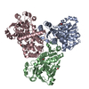
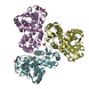
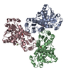
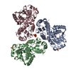
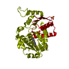
| Title | Aspartate transcarbamoylase domain of human CAD bound to carbamoyl phosphate | ||||||
 Components Components | CAD PROTEIN | ||||||
 Keywords Keywords | TRANSFERASE / DE NOVO PYRIMIDINE SYNTHESIS / TRANSCARBAMOYLASE / TRANSCARBAMYLASE / CAD / CARBAMOYL PHOSPHATE SYNTHETASE / DIHYDROOROTASE / COOPERATIVITY | ||||||
| Function / homology |  Function and homology information Function and homology informationaspartate binding / carbamoyl-phosphate synthase (glutamine-hydrolysing) / carbamoyl-phosphate synthase (ammonia) / carbamoyl-phosphate synthase (ammonia) activity / carbamoyl-phosphate synthase (glutamine-hydrolyzing) activity / dihydroorotase / L-citrulline biosynthetic process / response to cortisol / glutaminase / aspartate carbamoyltransferase ...aspartate binding / carbamoyl-phosphate synthase (glutamine-hydrolysing) / carbamoyl-phosphate synthase (ammonia) / carbamoyl-phosphate synthase (ammonia) activity / carbamoyl-phosphate synthase (glutamine-hydrolyzing) activity / dihydroorotase / L-citrulline biosynthetic process / response to cortisol / glutaminase / aspartate carbamoyltransferase / aspartate carbamoyltransferase activity / dihydroorotase activity / glutaminase activity / L-glutamine metabolic process / Pyrimidine biosynthesis / UTP biosynthetic process / UDP biosynthetic process / response to amine / response to caffeine / response to starvation / response to testosterone / animal organ regeneration / 'de novo' UMP biosynthetic process / 'de novo' pyrimidine nucleobase biosynthetic process / cell projection / lactation / xenobiotic metabolic process / cellular response to epidermal growth factor stimulus / liver development / female pregnancy / response to insulin / nuclear matrix / terminal bouton / heart development / protein kinase activity / neuronal cell body / enzyme binding / protein-containing complex / extracellular exosome / zinc ion binding / ATP binding / membrane / identical protein binding / nucleus / cytosol / cytoplasm Similarity search - Function | ||||||
| Biological species |  HOMO SAPIENS (human) HOMO SAPIENS (human) | ||||||
| Method |  X-RAY DIFFRACTION / SYNCHROTRON / MOLECULAR REPLACEMENT / Resolution: 3.19 Å X-RAY DIFFRACTION / SYNCHROTRON / MOLECULAR REPLACEMENT / Resolution: 3.19 Å | ||||||
 Authors Authors | Ruiz-Ramos, A. / Grande-Garcia, A. / Moreno-Morcillo, M.D. / Ramon-Maiques, S. | ||||||
 Citation Citation | Journal: Structure / Year: 2016 Title: Structure and Functional Characterization of Human Aspartate Transcarbamoylase, the Target of the Anti-Tumoral Drug Pala. Authors: Ruiz-Ramos, A. / Velazquez-Campoy, A. / Grande-Garcia, A. / Moreno-Morcillo, M. / Ramon-Maiques, S. #1: Journal: Acta Crystallogr.,Sect.F / Year: 2013 Title: Expression, Purification, Crystallization and Preliminary X- Ray Diffraction Analysis of the Aspartate Transcarbamoylase Domain of Human Cad. Authors: Ruiz-Ramos, A. / Lallous, N. / Grande-Garcia, A. / Ramon-Maiques, S. | ||||||
| History |
|
- Structure visualization
| Structure viewer | Molecule: MolmilJmol/JSmol |
|---|
- Downloads & links
-Download
| PDBx/mmCIF format | 5g1p.cif.gz | 335 KB | Display | PDBx/mmCIF format |
|---|---|---|---|---|
| PDB format | pdb5g1p.ent.gz | 270.6 KB | Display | PDB format |
| PDBx/mmJSON format | 5g1p.json.gz | Tree view | PDBx/mmJSON format | |
| Others |  Other downloads Other downloads |
-Validation report
| Arichive directory | https://data.pdbj.org/pub/pdb/validation_reports/g1/5g1pftp://data.pdbj.org/pub/pdb/validation_reports/g1/5g1p | HTTPS FTP |
|---|
-Related structure data
| Related structure data | 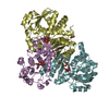 5g1nC 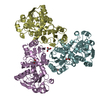 5g1oC  3csuS C: citing same article ( S: Starting model for refinement |
|---|---|
| Similar structure data |

-Links
 PDBj
PDBj


- Assembly
| Deposited unit | 
| ||||||||
|---|---|---|---|---|---|---|---|---|---|
| 1 | 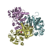
| ||||||||
| 2 | 
| ||||||||
| Unit cell |
|
-Components
| #1: Protein | Mass: 34911.422 Da / Num. of mol.: 6 Source method: isolated from a genetically manipulated source Source: (gene. exp.) HOMO SAPIENS (human)Description: HUMAN CDNA PURCHASED FROM OPENBIOSYSTEMS (CLONE ID 5551082) Plasmid: POPIN-M / Production host:  #2: Chemical | ChemComp-CP /   Mass: 141.020 Da / Num. of mol.: 6 / Source method: obtained synthetically / Formula: CH4NO5P Mass: 141.020 Da / Num. of mol.: 6 / Source method: obtained synthetically / Formula: CH4NO5P#3: Water | ChemComp-HOH / |  Mass: 18.015 Da / Num. of mol.: 95 / Source method: isolated from a natural source / Formula: H2O Mass: 18.015 Da / Num. of mol.: 95 / Source method: isolated from a natural source / Formula: H2O |
|---|
-Experimental details
-Experiment
| Experiment | Method: X-RAY DIFFRACTION / Number of used crystals: 1 |
|---|
- Sample preparation
| Crystal | Density Matthews: 2.1 Å3/Da / Density % sol: 41 % / Description: NONE |
|---|---|
| Crystal grow | Details: 0.1 M TRIS PH 8.5, 6% PEG 8000, 11% ETHYLENE GLYCOL |
-Data collection
| Diffraction | Mean temperature: 100 K | |||||||||||||||||||||||||||||||||||
|---|---|---|---|---|---|---|---|---|---|---|---|---|---|---|---|---|---|---|---|---|---|---|---|---|---|---|---|---|---|---|---|---|---|---|---|---|
| Diffraction source | Source: SYNCHROTRON / Site: SLS  / Beamline: X06SA / Wavelength: 0.976 / Beamline: X06SA / Wavelength: 0.976 | |||||||||||||||||||||||||||||||||||
| Detector | Type: DECTRIS PILATUS 6M / Detector: PIXEL / Date: May 9, 2013 | |||||||||||||||||||||||||||||||||||
| Radiation | Protocol: SINGLE WAVELENGTH / Monochromatic (M) / Laue (L): M / Scattering type: x-ray | |||||||||||||||||||||||||||||||||||
| Radiation wavelength | Wavelength: 0.976 Å / Relative weight: 1 | |||||||||||||||||||||||||||||||||||
| Reflection twin |
| |||||||||||||||||||||||||||||||||||
| Reflection | Resolution: 3.2→50 Å / Num. obs: 29277 / % possible obs: 99.4 % / Observed criterion σ(I): 1 / Redundancy: 4.8 % / Rmerge(I) obs: 0.17 / Net I/σ(I): 9.9 | |||||||||||||||||||||||||||||||||||
| Reflection shell | Resolution: 3.2→3.28 Å / Redundancy: 4.9 % / Rmerge(I) obs: 0.78 / Mean I/σ(I) obs: 2.3 / % possible all: 99.9 |
- Processing
| Software |
| ||||||||||||||||||||||||||||||||||||||||||||||||||||||||||||||||||||||||||||||||||||||||||||||||||||||||||||||||||||||||||||||||||||||||||||||||||||||||||||||||||||||||||||||||||||||
|---|---|---|---|---|---|---|---|---|---|---|---|---|---|---|---|---|---|---|---|---|---|---|---|---|---|---|---|---|---|---|---|---|---|---|---|---|---|---|---|---|---|---|---|---|---|---|---|---|---|---|---|---|---|---|---|---|---|---|---|---|---|---|---|---|---|---|---|---|---|---|---|---|---|---|---|---|---|---|---|---|---|---|---|---|---|---|---|---|---|---|---|---|---|---|---|---|---|---|---|---|---|---|---|---|---|---|---|---|---|---|---|---|---|---|---|---|---|---|---|---|---|---|---|---|---|---|---|---|---|---|---|---|---|---|---|---|---|---|---|---|---|---|---|---|---|---|---|---|---|---|---|---|---|---|---|---|---|---|---|---|---|---|---|---|---|---|---|---|---|---|---|---|---|---|---|---|---|---|---|---|---|---|---|
| Refinement | Method to determine structure: MOLECULAR REPLACEMENT Starting model: PDB ENTRY 3CSU Resolution: 3.19→42.54 Å / Cor.coef. Fo:Fc: 0.858 / Cor.coef. Fo:Fc free: 0.831 / SU B: 19.999 / SU ML: 0.383 / Cross valid method: THROUGHOUT / ESU R Free: 0.116 / Stereochemistry target values: MAXIMUM LIKELIHOOD / Details: HYDROGENS HAVE BEEN ADDED IN THE RIDING POSITIONS.
| ||||||||||||||||||||||||||||||||||||||||||||||||||||||||||||||||||||||||||||||||||||||||||||||||||||||||||||||||||||||||||||||||||||||||||||||||||||||||||||||||||||||||||||||||||||||
| Solvent computation | Ion probe radii: 0.8 Å / Shrinkage radii: 0.8 Å / VDW probe radii: 1.2 Å / Solvent model: MASK | ||||||||||||||||||||||||||||||||||||||||||||||||||||||||||||||||||||||||||||||||||||||||||||||||||||||||||||||||||||||||||||||||||||||||||||||||||||||||||||||||||||||||||||||||||||||
| Displacement parameters | Biso mean: 50.033 Å2
| ||||||||||||||||||||||||||||||||||||||||||||||||||||||||||||||||||||||||||||||||||||||||||||||||||||||||||||||||||||||||||||||||||||||||||||||||||||||||||||||||||||||||||||||||||||||
| Refinement step | Cycle: LAST / Resolution: 3.19→42.54 Å
| ||||||||||||||||||||||||||||||||||||||||||||||||||||||||||||||||||||||||||||||||||||||||||||||||||||||||||||||||||||||||||||||||||||||||||||||||||||||||||||||||||||||||||||||||||||||
| Refine LS restraints |
|