 Movie
Movie Controller

[English] 日本語
 Yorodumi
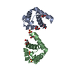
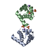
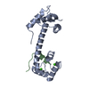
- PDB-1ahr: CALMODULIN MUTANT WITH A TWO RESIDUE DELETION IN THE CENTRAL HELIX -
+ Open data

- Basic information
| Entry | Database: PDB / ID: 1ahr | ||||||
|---|---|---|---|---|---|---|---|
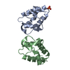
| Title | CALMODULIN MUTANT WITH A TWO RESIDUE DELETION IN THE CENTRAL HELIX | ||||||
 Components Components | CALMODULIN | ||||||
 Keywords Keywords | CALCIUM-BINDING PROTEIN / CALMODULIN | ||||||
| Function / homology |  Function and homology information Function and homology informationCH domain binding / myosin binding / calcineurin-mediated signaling / detection of calcium ion / regulation of release of sequestered calcium ion into cytosol by sarcoplasmic reticulum / disordered domain specific binding / myelin sheath / synaptic vesicle membrane / calcium ion binding / centrosome ...CH domain binding / myosin binding / calcineurin-mediated signaling / detection of calcium ion / regulation of release of sequestered calcium ion into cytosol by sarcoplasmic reticulum / disordered domain specific binding / myelin sheath / synaptic vesicle membrane / calcium ion binding / centrosome / protein-containing complex / cytoplasm Similarity search - Function | ||||||
| Biological species |  | ||||||
| Method |  X-RAY DIFFRACTION / MOLECULAR REPLACEMENT / Resolution: 1.8 Å X-RAY DIFFRACTION / MOLECULAR REPLACEMENT / Resolution: 1.8 Å | ||||||
 Authors Authors | Tabernero, L. / Sack, J. | ||||||
 Citation Citation | Journal: Structure / Year: 1997 Title: The structure of a calmodulin mutant with a deletion in the central helix: implications for molecular recognition and protein binding. Authors: Tabernero, L. / Taylor, D.A. / Chandross, R.J. / VanBerkum, M.F. / Means, A.R. / Quiocho, F.A. / Sack, J.S. | ||||||
| History |
|
- Structure visualization
| Structure viewer | Molecule: MolmilJmol/JSmol |
|---|
- Downloads & links
-Download
| PDBx/mmCIF format | 1ahr.cif.gz | 44.1 KB | Display | PDBx/mmCIF format |
|---|---|---|---|---|
| PDB format | pdb1ahr.ent.gz | 30.8 KB | Display | PDB format |
| PDBx/mmJSON format | 1ahr.json.gz | Tree view | PDBx/mmJSON format | |
| Others |  Other downloads Other downloads |
-Validation report
| Arichive directory | https://data.pdbj.org/pub/pdb/validation_reports/ah/1ahrftp://data.pdbj.org/pub/pdb/validation_reports/ah/1ahr | HTTPS FTP |
|---|
-Related structure data
| Related structure data | 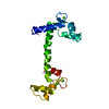 4clnS S: Starting model for refinement |
|---|---|
| Similar structure data |

-Links
 PDBj
PDBj

- Assembly
| Deposited unit | 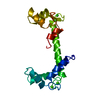
| ||||||||
|---|---|---|---|---|---|---|---|---|---|
| 1 |
| ||||||||
| Unit cell |
|
-Components
| #1: Protein | Mass: 16478.133 Da / Num. of mol.: 1 / Mutation: DEL(T79), DEL(D80) Source method: isolated from a genetically manipulated source Source: (gene. exp.)  | ||||
|---|---|---|---|---|---|
| #2: Chemical | ChemComp-CA /   Mass: 40.078 Da / Num. of mol.: 4 / Source method: obtained synthetically / Formula: Ca Mass: 40.078 Da / Num. of mol.: 4 / Source method: obtained synthetically / Formula: Ca#3: Water | ChemComp-HOH / |  Mass: 18.015 Da / Num. of mol.: 113 / Source method: isolated from a natural source / Formula: H2O Mass: 18.015 Da / Num. of mol.: 113 / Source method: isolated from a natural source / Formula: H2OSequence details | SEQUENCE AND NUMBERING IS AS IN WILD TYPE CALMODULIN | |
-Experimental details
-Experiment
| Experiment | Method: X-RAY DIFFRACTION / Number of used crystals: 1 |
|---|
- Sample preparation
| Crystal | Density Matthews: 2.32 Å3/Da / Density % sol: 46.56 % / Description: SEPARATE ROTATION SEARCH FOR EACH DOMAIN | ||||||||||||||||||||||||||||||
|---|---|---|---|---|---|---|---|---|---|---|---|---|---|---|---|---|---|---|---|---|---|---|---|---|---|---|---|---|---|---|---|
| Crystal grow | pH: 4.25 Details: 28-33% MPD, 15% ETHANOL, 5MM CACL, 20MM NA ACETATE, PH 4.25 | ||||||||||||||||||||||||||||||
| Crystal grow | *PLUS Method: vapor diffusion, hanging drop | ||||||||||||||||||||||||||||||
| Components of the solutions | *PLUS
|
-Data collection
| Diffraction | Mean temperature: 300 K |
|---|---|
| Diffraction source | Source: ROTATING ANODE / Type: RIGAKU RUH2R / Wavelength: 1.5418 |
| Detector | Type: ADSC / Detector: AREA DETECTOR / Date: Dec 1, 1988 / Details: COLLIMATOR |
| Radiation | Monochromator: GRAPHITE(002) / Monochromatic (M) / Laue (L): M / Scattering type: x-ray |
| Radiation wavelength | Wavelength: 1.5418 Å / Relative weight: 1 |
| Reflection | Highest resolution: 1.8 Å / Num. obs: 14407 / % possible obs: 67.3 % / Observed criterion σ(I): 2 / Redundancy: 2.4 % / Rsym value: 0.501 / Net I/σ(I): 8.68 |
| Reflection | *PLUS Num. measured all: 27799 / Rmerge(I) obs: 0.0501 |
- Processing
| Software |
| ||||||||||||||||||||||||||||||||||||||||||||||||||||||||||||
|---|---|---|---|---|---|---|---|---|---|---|---|---|---|---|---|---|---|---|---|---|---|---|---|---|---|---|---|---|---|---|---|---|---|---|---|---|---|---|---|---|---|---|---|---|---|---|---|---|---|---|---|---|---|---|---|---|---|---|---|---|---|
| Refinement | Method to determine structure: MOLECULAR REPLACEMENT Starting model: PDB ENTRY 4CLN Resolution: 1.8→8 Å / Rfactor Rwork: 0.211 / Rfactor obs: 0.211 / σ(F): 2 | ||||||||||||||||||||||||||||||||||||||||||||||||||||||||||||
| Refinement step | Cycle: LAST / Resolution: 1.8→8 Å
| ||||||||||||||||||||||||||||||||||||||||||||||||||||||||||||
| Refine LS restraints |
| ||||||||||||||||||||||||||||||||||||||||||||||||||||||||||||
| Software | *PLUS Name: X-PLOR / Version: 3.1 / Classification: refinement | ||||||||||||||||||||||||||||||||||||||||||||||||||||||||||||
| Refinement | *PLUS Num. reflection obs: 9845 | ||||||||||||||||||||||||||||||||||||||||||||||||||||||||||||
| Solvent computation | *PLUS | ||||||||||||||||||||||||||||||||||||||||||||||||||||||||||||
| Displacement parameters | *PLUS | ||||||||||||||||||||||||||||||||||||||||||||||||||||||||||||
| Refine LS restraints | *PLUS Type: x_bond_d / Dev ideal: 0.015 |
