| Entry | Database: PDB / ID: 4xuw
|
|---|
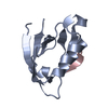
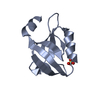
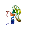
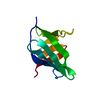
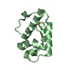
| Title | Structure of the hazelnut allergen, Cor a 8 |
|---|
 Components Components | Non-specific lipid-transfer protein |
|---|
 Keywords Keywords | ALLERGEN / plant protein |
|---|
| Function / homology |  Function and homology information Function and homology information
Plant lipid transfer proteins signature. / Plant non-specific lipid-transfer protein/Par allergen / Plant lipid-transfer and hydrophobic proteins / Hydrophobic Seed Protein / Protease inhibitor/seed storage/LTP family / Plant lipid transfer protein / seed storage protein / trypsin-alpha amylase inhibitor domain family / Bifunctional inhibitor/plant lipid transfer protein/seed storage helical domain / Bifunctional inhibitor/plant lipid transfer protein/seed storage helical domain superfamily / Orthogonal Bundle / Mainly AlphaSimilarity search - Domain/homology |
|---|
| Biological species |  Corylus avellana (European hazelnut) Corylus avellana (European hazelnut) |
|---|
| Method |  X-RAY DIFFRACTION / SYNCHROTRON / MOLECULAR REPLACEMENT / Resolution: 1.1 Å X-RAY DIFFRACTION / SYNCHROTRON / MOLECULAR REPLACEMENT / Resolution: 1.1 Å |
|---|
 Authors Authors | Offermann, L.R. / Perdue, M.L. / Bublin, M. / Pfeifer, S. / Dubiela, P. / Hoffmann-Sommergruber, K. / Chruszcz, M. |
|---|
 Citation Citation | Journal: J.Agric.Food Chem. / Year: 2015
Title: Structural and Functional Characterization of the Hazelnut Allergen Cor a 8.
Authors: Offermann, L.R. / Bublin, M. / Perdue, M.L. / Pfeifer, S. / Dubiela, P. / Borowski, T. / Chruszcz, M. / Hoffmann-Sommergruber, K. |
|---|
| History | | Deposition | Jan 26, 2015 | Deposition site: RCSB / Processing site: RCSB |
|---|
| Revision 1.0 | Oct 14, 2015 | Provider: repository / Type: Initial release |
|---|
| Revision 1.1 | Nov 4, 2015 | Group: Database references |
|---|
| Revision 1.2 | Sep 27, 2023 | Group: Data collection / Database references ...Data collection / Database references / Derived calculations / Refinement description
Category: chem_comp_atom / chem_comp_bond ...chem_comp_atom / chem_comp_bond / database_2 / pdbx_initial_refinement_model / pdbx_struct_oper_list / struct_ncs_dom_lim
Item: _database_2.pdbx_DOI / _database_2.pdbx_database_accession ..._database_2.pdbx_DOI / _database_2.pdbx_database_accession / _pdbx_struct_oper_list.symmetry_operation / _struct_ncs_dom_lim.beg_auth_comp_id / _struct_ncs_dom_lim.beg_label_asym_id / _struct_ncs_dom_lim.beg_label_comp_id / _struct_ncs_dom_lim.beg_label_seq_id / _struct_ncs_dom_lim.end_auth_comp_id / _struct_ncs_dom_lim.end_label_asym_id / _struct_ncs_dom_lim.end_label_comp_id / _struct_ncs_dom_lim.end_label_seq_id |
|---|
| Revision 1.3 | Oct 16, 2024 | Group: Structure summary / Category: pdbx_entry_details / pdbx_modification_feature |
|---|
|
|---|
 Movie
Movie Controller


 Open data
 Basic information
 Structure visualization
 Downloads & links
 Other downloads
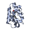


 PDBj
PDBj
 Assembly
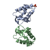


 Mass: 94.971 Da / Num. of mol.: 1 / Source method: obtained synthetically / Formula: PO4
 Mass: 18.015 Da / Num. of mol.: 308 / Source method: isolated from a natural source / Formula: H2O
 Sample preparation
 / Beamline: 22-BM / Wavelength: 1 Å
 Processing