toxin-antitoxin complex / single-species biofilm formation / sequence-specific DNA binding / regulation of DNA-templated transcription / protein homodimerization activity / metal ion binding Similarity search - Function
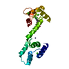
Method: simulated annealing / Software ordinal: 1 Details: 3 7 CYCLES OF CYANA - CANDID. A) KEEPING MANUAL ASSIGNEMENTS B) ALLOWING MANUAL ASSIGNEMENTS TO VARIATE C) ADDING EXTRA CSVALUES FROM STATISTICAL TABLES. IF THE N-TERMINUS DOMAIN IS ALIGNED ...Details: 3 7 CYCLES OF CYANA - CANDID. A) KEEPING MANUAL ASSIGNEMENTS B) ALLOWING MANUAL ASSIGNEMENTS TO VARIATE C) ADDING EXTRA CSVALUES FROM STATISTICAL TABLES. IF THE N-TERMINUS DOMAIN IS ALIGNED IT LOOKS STRUCTURED AND THE C-TERMINUS ROTATES. THE TWO ALIGNMENTS GIVE A SOMEHOW DIFFERENT IMPRESSION ABOUT THE PROTEIN AND VERIFY THAT EACH DOMAIN IS INDIVIDUALLY STRUCTURED. THERE ARE TWO B-SHEETS ONE WITH 4 STRANDS AND ONE WITH TWO LONG STRANDS. THE 4 STRANDS MIGHT BELONG TO THE SAME SHEET OR THEY MAY BE TWO TIMES TWO STRANDS OR A SMALL B-BARREL. THEY BIND AND THEY ARE STABILIZED BY ZN.
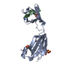
NMR representative
Selection criteria: fewest violations
NMR ensemble
Conformer selection criteria: structures with acceptable covalent geometry Conformers calculated total number: 100 / Conformers submitted total number: 20 / Representative conformer: 1
+
About Yorodumi
-
News
-
Feb 9, 2022. New format data for meta-information of EMDB entries
New format data for meta-information of EMDB entries
Version 3 of the EMDB header file is now the official format.
The previous official version 1.9 will be removed from the archive.
In the structure databanks used in Yorodumi, some data are registered as the other names, "COVID-19 virus" and "2019-nCoV". Here are the details of the virus and the list of structure data.
Jan 31, 2019. EMDB accession codes are about to change! (news from PDBe EMDB page)
EMDB accession codes are about to change! (news from PDBe EMDB page)
The allocation of 4 digits for EMDB accession codes will soon come to an end. Whilst these codes will remain in use, new EMDB accession codes will include an additional digit and will expand incrementally as the available range of codes is exhausted. The current 4-digit format prefixed with “EMD-” (i.e. EMD-XXXX) will advance to a 5-digit format (i.e. EMD-XXXXX), and so on. It is currently estimated that the 4-digit codes will be depleted around Spring 2019, at which point the 5-digit format will come into force.
The EM Navigator/Yorodumi systems omit the EMD- prefix.
Related info.:Q: What is EMD? / ID/Accession-code notation in Yorodumi/EM Navigator
Yorodumi is a browser for structure data from EMDB, PDB, SASBDB, etc.
This page is also the successor to EM Navigator detail page, and also detail information page/front-end page for Omokage search.
The word "yorodu" (or yorozu) is an old Japanese word meaning "ten thousand". "mi" (miru) is to see.
Related info.:EMDB / PDB / SASBDB / Comparison of 3 databanks / Yorodumi Search / Aug 31, 2016. New EM Navigator & Yorodumi / Yorodumi Papers / Jmol/JSmol / Function and homology information / Changes in new EM Navigator and Yorodumi
 Movie
Movie Controller

 Yorodumi
 Open data
 Basic information
 Components
 Keywords
 Function and homology information
 Authors
 Citation
 Structure visualization
 Downloads & links
 Other downloads
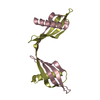


 PDBj
PDBj


 Assembly
 HSQC
HSQC Sample preparation
 Processing