 Movie
Movie Controller


+ Open data

- Basic information
| Entry | Database: PDB / ID: 1adu | ||||||
|---|---|---|---|---|---|---|---|
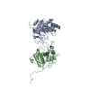
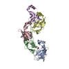
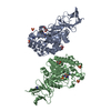
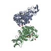
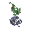
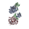
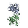
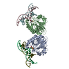
| Title | EARLY E2A DNA-BINDING PROTEIN | ||||||
 Components Components | ADENOVIRUS SINGLE-STRANDED DNA-BINDING PROTEIN | ||||||
 Keywords Keywords | DNA BINDING PROTEIN / DNA-BINDING PROTEIN / SSDNA BINDING PROTEIN | ||||||
| Function / homology |  Function and homology information Function and homology informationviral DNA strand displacement replication / viral DNA genome replication / positive regulation of DNA replication / viral capsid / single-stranded DNA binding / DNA replication / DNA-templated transcription / host cell nucleus / DNA binding / zinc ion binding / identical protein binding Similarity search - Function | ||||||
| Biological species |   Human adenovirus 5 Human adenovirus 5 | ||||||
| Method |  X-RAY DIFFRACTION / SYNCHROTRON / Resolution: 3 Å X-RAY DIFFRACTION / SYNCHROTRON / Resolution: 3 Å | ||||||
 Authors Authors | Tucker, P.A. / Kanellopoulos, P.N. / Tsernoglou, D. / Van Der Vliet, P.C. | ||||||
 Citation Citation | Journal: J.Mol.Biol. / Year: 1996 Title: Alternative arrangements of the protein chain are possible for the adenovirus single-stranded DNA binding protein. Authors: Kanellopoulos, P.N. / Tsernoglou, D. / van der Vliet, P.C. / Tucker, P.A. #1: Journal: Embo J. / Year: 1994Title: Crystal Structure of the Adenovirus DNA Binding Protein Reveals a Hook-on Model for Cooperative DNA Binding Authors: Tucker, P.A. / Tsernoglou, D. / Tucker, A.D. / Coenjaerts, F.E. / Leenders, H. / Van Der Vliet, P.C. | ||||||
| History |
|
- Structure visualization
| Structure viewer | Molecule: MolmilJmol/JSmol |
|---|
- Downloads & links
-Download
| PDBx/mmCIF format | 1adu.cif.gz | 124.2 KB | Display | PDBx/mmCIF format |
|---|---|---|---|---|
| PDB format | pdb1adu.ent.gz | 96.5 KB | Display | PDB format |
| PDBx/mmJSON format | 1adu.json.gz | Tree view | PDBx/mmJSON format | |
| Others |  Other downloads Other downloads |
-Validation report
| Arichive directory | https://data.pdbj.org/pub/pdb/validation_reports/ad/1aduftp://data.pdbj.org/pub/pdb/validation_reports/ad/1adu | HTTPS FTP |
|---|
-Related structure data

-Links
 PDBj
PDBj- Assembly
| Deposited unit | 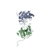
| ||||||||
|---|---|---|---|---|---|---|---|---|---|
| 1 |
| ||||||||
| Unit cell |
| ||||||||
| Details | THE MOLECULES FORM CHAINS ALONG THE CRYSTALLOGRAPHIC Z AXIS. PAIRS OF MOLECULES IN THE CHAIN ARE RELATED BY A CRYSTALLOGRAPHIC SCREW AXIS. |
-Components
| #1: Protein | Mass: 39899.746 Da / Num. of mol.: 2 / Fragment: C-TERMINAL DOMAIN, RESIDUES 174 - 529 / Source method: isolated from a natural source / Details: SECOND CRYSTAL FORM, DATA COLLECTED AT 293 K / Source: (natural) Human adenovirus 5 / Genus: Mastadenovirus / Cell: INFECTED HELA CELLS / Species: Human adenovirus C / References: UniProt: P03265#2: Chemical | ChemComp-ZN /   Mass: 65.409 Da / Num. of mol.: 4 / Source method: obtained synthetically / Formula: Zn Mass: 65.409 Da / Num. of mol.: 4 / Source method: obtained synthetically / Formula: Zn |
|---|
-Experimental details
-Experiment
| Experiment | Method: X-RAY DIFFRACTION |
|---|
- Sample preparation
| Crystal | Density Matthews: 2.6 Å3/Da / Density % sol: 52.74 % | |||||||||||||||
|---|---|---|---|---|---|---|---|---|---|---|---|---|---|---|---|---|
| Crystal grow | *PLUS Method: vapor diffusion | |||||||||||||||
| Components of the solutions | *PLUS
|
-Data collection
| Diffraction | Mean temperature: 293 K |
|---|---|
| Diffraction source | Source: SYNCHROTRON / Site: EMBL/DESY, HAMBURG  / Beamline: BW7B / Wavelength: 0.97 / Beamline: BW7B / Wavelength: 0.97 |
| Detector | Type: MARRESEARCH / Detector: IMAGE PLATE / Date: Jun 27, 1994 |
| Radiation | Monochromatic (M) / Laue (L): M / Scattering type: x-ray |
| Radiation wavelength | Wavelength: 0.97 Å / Relative weight: 1 |
| Reflection | Resolution: 3→20 Å / Num. obs: 17172 / % possible obs: 99.5 % / Observed criterion σ(I): 0 / Rmerge(I) obs: 0.168 |
| Reflection | *PLUS Num. measured all: 73641 |
| Reflection shell | *PLUS Highest resolution: 3 Å / Lowest resolution: 3.2 Å / Rmerge(I) obs: 0.496 |
- Processing
| Software |
| ||||||||||||||||||||||||||||||||||||||||||||||||||||||||||||
|---|---|---|---|---|---|---|---|---|---|---|---|---|---|---|---|---|---|---|---|---|---|---|---|---|---|---|---|---|---|---|---|---|---|---|---|---|---|---|---|---|---|---|---|---|---|---|---|---|---|---|---|---|---|---|---|---|---|---|---|---|---|
| Refinement | Resolution: 3→6 Å / σ(F): 0
| ||||||||||||||||||||||||||||||||||||||||||||||||||||||||||||
| Displacement parameters | Biso mean: 35.45 Å2 | ||||||||||||||||||||||||||||||||||||||||||||||||||||||||||||
| Refine analyze | Luzzati coordinate error free: 0.35 Å | ||||||||||||||||||||||||||||||||||||||||||||||||||||||||||||
| Refinement step | Cycle: LAST / Resolution: 3→6 Å
| ||||||||||||||||||||||||||||||||||||||||||||||||||||||||||||
| Refine LS restraints |
| ||||||||||||||||||||||||||||||||||||||||||||||||||||||||||||
| Software | *PLUS Name: X-PLOR / Classification: refinement | ||||||||||||||||||||||||||||||||||||||||||||||||||||||||||||
| Refinement | *PLUS | ||||||||||||||||||||||||||||||||||||||||||||||||||||||||||||
| Solvent computation | *PLUS | ||||||||||||||||||||||||||||||||||||||||||||||||||||||||||||
| Displacement parameters | *PLUS | ||||||||||||||||||||||||||||||||||||||||||||||||||||||||||||
| Refine LS restraints | *PLUS
|
