 Movie
Movie Controller

[English] 日本語
 Yorodumi
- PDB-1adq: CRYSTAL STRUCTURE OF A HUMAN IGM RHEUMATOID FACTOR FAB IN COMPLEX... -
+ Open data

- Basic information
| Entry | Database: PDB / ID: 1adq | ||||||
|---|---|---|---|---|---|---|---|
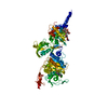
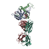
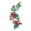
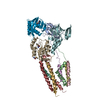
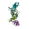
| Title | CRYSTAL STRUCTURE OF A HUMAN IGM RHEUMATOID FACTOR FAB IN COMPLEX WITH ITS AUTOANTIGEN IGG FC | ||||||
 Components Components |
| ||||||
 Keywords Keywords | COMPLEX (IMMUNOGLOBULIN/AUTOANTIGEN) / COMPLEX (IMMUNOGLOBULIN-AUTOANTIGEN) / RHEUMATOID FACTOR AUTO-ANTIBODY COMPLEX / COMPLEX (IMMUNOGLOBULIN-AUTOANTIGEN) complex | ||||||
| Function / homology |  Function and homology information Function and homology informationimmunoglobulin complex, circulating / Classical antibody-mediated complement activation / immunoglobulin receptor binding / Initial triggering of complement / FCGR activation / complement activation, classical pathway / Role of phospholipids in phagocytosis / antigen binding / FCGR3A-mediated IL10 synthesis / Regulation of Complement cascade ...immunoglobulin complex, circulating / Classical antibody-mediated complement activation / immunoglobulin receptor binding / Initial triggering of complement / FCGR activation / complement activation, classical pathway / Role of phospholipids in phagocytosis / antigen binding / FCGR3A-mediated IL10 synthesis / Regulation of Complement cascade / B cell receptor signaling pathway / FCGR3A-mediated phagocytosis / Regulation of actin dynamics for phagocytic cup formation / antibacterial humoral response / Interleukin-4 and Interleukin-13 signaling / blood microparticle / adaptive immune response / : / extracellular exosome / extracellular region / plasma membrane Similarity search - Function | ||||||
| Biological species |  Homo sapiens (human) Homo sapiens (human) | ||||||
| Method |  X-RAY DIFFRACTION / MOLECULAR REPLACEMENT / Resolution: 3.15 Å X-RAY DIFFRACTION / MOLECULAR REPLACEMENT / Resolution: 3.15 Å | ||||||
 Authors Authors | Corper, A.L. / Taussig, M.J. / Sutton, B.J. | ||||||
 Citation Citation | Journal: Nat.Struct.Biol. / Year: 1997 Title: Structure of human IgM rheumatoid factor Fab bound to its autoantigen IgG Fc reveals a novel topology of antibody-antigen interaction. Authors: Corper, A.L. / Sohi, M.K. / Bonagura, V.R. / Steinitz, M. / Jefferis, R. / Feinstein, A. / Beale, D. / Taussig, M.J. / Sutton, B.J. #1: Journal: Immunology / Year: 1996Title: Crystallization of a Complex between the Fab Fragment of a Human Immunoglobulin M (Igm) Rheumatoid Factor (Rf-an) and the Fc Fragment of Human Igg4 Authors: Sohi, M.K. / Corper, A.L. / Wan, T. / Steinitz, M. / Jefferis, R. / Beale, D. / He, M. / Feinstein, A. / Sutton, B.J. / Taussig, M.J. | ||||||
| History |
|
- Structure visualization
| Structure viewer | Molecule: MolmilJmol/JSmol |
|---|
- Downloads & links
-Download
| PDBx/mmCIF format | 1adq.cif.gz | 126.1 KB | Display | PDBx/mmCIF format |
|---|---|---|---|---|
| PDB format | pdb1adq.ent.gz | 94.7 KB | Display | PDB format |
| PDBx/mmJSON format | 1adq.json.gz | Tree view | PDBx/mmJSON format | |
| Others |  Other downloads Other downloads |
-Validation report
| Arichive directory | https://data.pdbj.org/pub/pdb/validation_reports/ad/1adqftp://data.pdbj.org/pub/pdb/validation_reports/ad/1adq | HTTPS FTP |
|---|
-Related structure data


-Links
 PDBj
PDBj


- Assembly
| Deposited unit | 
| ||||||||
|---|---|---|---|---|---|---|---|---|---|
| 1 |
| ||||||||
| Unit cell |
|
-Components
| #1: Protein | Mass: 23484.457 Da / Num. of mol.: 1 / Fragment: FC / Source method: isolated from a natural source Details: ISOLATED FROM THE SERA OF PATIENTS WITH MULTIPLE MYELOMA Source: (natural) Homo sapiens (human) / Cell: B-LYMPHOCYTE / Cellular location: EXTRACELLULAR / References: UniProt: P01861 |
|---|---|
| #2: Antibody | Mass: 22567.885 Da / Num. of mol.: 1 / Fragment: FAB / Source method: isolated from a natural source Details: RF-AN CELL LINE WAS PREPARED FROM PERIPHERAL BLOOD LYMPHOCYTES OF AN RA PATIENT BY TRANSFORMATION WITH EPSTEIN-BARR VIRUS Source: (natural) Homo sapiens (human) / Cell: B-LYMPHOCYTE / Cell line: RF-AN / Cellular location: EXTRACELLULAR |
| #3: Antibody | Mass: 24665.742 Da / Num. of mol.: 1 / Fragment: FAB / Source method: isolated from a natural source Details: RF-AN CELL LINE WAS PREPARED FROM PERIPHERAL BLOOD LYMPHOCYTES OF AN RA PATIENT BY TRANSFORMATION WITH EPSTEIN-BARR VIRUS Source: (natural) Homo sapiens (human) / Cell: B-LYMPHOCYTE / Cell line: RF-AN / Cellular location: EXTRACELLULAR |
| Has protein modification | Y |
-Experimental details
-Experiment
| Experiment | Method: X-RAY DIFFRACTION / Number of used crystals: 1 |
|---|
- Sample preparation
| Crystal | Density Matthews: 2.9 Å3/Da / Density % sol: 58 % | ||||||||||||||||||||||||||||||||||||||||||
|---|---|---|---|---|---|---|---|---|---|---|---|---|---|---|---|---|---|---|---|---|---|---|---|---|---|---|---|---|---|---|---|---|---|---|---|---|---|---|---|---|---|---|---|
| Crystal grow | Method: vapor diffusion, hanging drop / pH: 7 Details: CRYSTALS WERE GROWN BY HANGING DROP VAPOR DIFFUSION. THE HANGING DROPS CONSISTED OF 2UL OF PROTEIN SOLUTION CONTAINING EACH PROTEIN AT 1MG/ML IN 0.1 % SODIUM AZIDE, 20MM TRIS-HCL AT PH 7.0, ...Details: CRYSTALS WERE GROWN BY HANGING DROP VAPOR DIFFUSION. THE HANGING DROPS CONSISTED OF 2UL OF PROTEIN SOLUTION CONTAINING EACH PROTEIN AT 1MG/ML IN 0.1 % SODIUM AZIDE, 20MM TRIS-HCL AT PH 7.0, MIXED WITH AN EQUAL VOLUME OF THE RESERVOIR SOLUTION TO BE SCREENED. CRYSTALS WERE OBTAINED WITH RESERVOIR SOLUTIONS CONTAINING 17.5 - 22.5 % (W/V) POLYETHYLENE GLYCOL (MEAN MW 8000) IN 0.1 % SODIUM AZIDE, 100MM TRIS-HCL, PH 7.0, AT TEMPERATURES BETWEEN 17.5 AND 21.5 (CELSIUS)., vapor diffusion - hanging drop PH range: APPROX. / Temp details: 290.5 - 294.5 | ||||||||||||||||||||||||||||||||||||||||||
| Crystal grow | *PLUS Temperature: 17.5-21.5 ℃ / Method: vapor diffusion, hanging dropDetails: drop consists of equal volume of protein and reservoir solutions | ||||||||||||||||||||||||||||||||||||||||||
| Components of the solutions | *PLUS
|
-Data collection
| Diffraction | Mean temperature: 278 K |
|---|---|
| Diffraction source | Source: ROTATING ANODE / Type: RIGAKU RU200 / Wavelength: 1.5418 |
| Detector | Type: RIGAKU RAXIS II / Detector: IMAGE PLATE / Date: Jun 1, 1994 / Details: COLLIMATOR |
| Radiation | Monochromator: GRAPHITE / Monochromatic (M) / Laue (L): M / Scattering type: x-ray |
| Radiation wavelength | Wavelength: 1.5418 Å / Relative weight: 1 |
| Reflection | Resolution: 3.15→15 Å / Num. obs: 14109 / % possible obs: 98.3 % / Observed criterion σ(I): 3 / Redundancy: 3.1 % / Biso Wilson estimate: 25.4 Å2 / Rmerge(I) obs: 0.113 / Net I/σ(I): 5.3 |
| Reflection shell | Resolution: 3.15→3.31 Å / Redundancy: 3.1 % / Rmerge(I) obs: 0.265 / Mean I/σ(I) obs: 2.8 / % possible all: 98.8 |
| Reflection shell | *PLUS % possible obs: 98.8 % |
- Processing
| Software |
| ||||||||||||||||||||||||||||||||||||||||||||||||||||||||||||
|---|---|---|---|---|---|---|---|---|---|---|---|---|---|---|---|---|---|---|---|---|---|---|---|---|---|---|---|---|---|---|---|---|---|---|---|---|---|---|---|---|---|---|---|---|---|---|---|---|---|---|---|---|---|---|---|---|---|---|---|---|---|
| Refinement | Method to determine structure: MOLECULAR REPLACEMENT Starting model: PDB ENTRY 1FC1, 2IG2 Resolution: 3.15→8 Å / Rfactor Rfree error: 0.011 / Data cutoff high absF: 100000 / Data cutoff low absF: 80 / Isotropic thermal model: GROUPED B-FACTOR REFINEMENT / Cross valid method: THROUGHOUT / σ(F): 0 Details: DISORDERED REGION A263-A300 WAS MODELED STEREOCHEMICALLY. FAB IN COMPLEX WITH ITS AUTOANTIGEN IGG FC. CONSTANT DOMAINS OF RF-AN SHOWED SIGNIFICANT DISORDER AND WERE PARTLY MODELED ...Details: DISORDERED REGION A263-A300 WAS MODELED STEREOCHEMICALLY. FAB IN COMPLEX WITH ITS AUTOANTIGEN IGG FC. CONSTANT DOMAINS OF RF-AN SHOWED SIGNIFICANT DISORDER AND WERE PARTLY MODELED STEREOCHEMICALLY. RESIDUES L51 AND L171 OCCUR IN WELL DEFINED REGIONS OF THE STRUCTURE (OCCUPANCY = 1). BOTH OCCUR IN LOOP REGIONS AND CONTINUE TO HAVE DISALLOWED PHI/PSI VALUES EVEN AFTER REBUILDING.
| ||||||||||||||||||||||||||||||||||||||||||||||||||||||||||||
| Displacement parameters | Biso mean: 33.1 Å2 | ||||||||||||||||||||||||||||||||||||||||||||||||||||||||||||
| Refine analyze |
| ||||||||||||||||||||||||||||||||||||||||||||||||||||||||||||
| Refinement step | Cycle: LAST / Resolution: 3.15→8 Å
| ||||||||||||||||||||||||||||||||||||||||||||||||||||||||||||
| Refine LS restraints |
| ||||||||||||||||||||||||||||||||||||||||||||||||||||||||||||
| LS refinement shell | Resolution: 3.15→3.26 Å / Rfactor Rfree error: 0.05 / Total num. of bins used: 10
| ||||||||||||||||||||||||||||||||||||||||||||||||||||||||||||
| Xplor file |
| ||||||||||||||||||||||||||||||||||||||||||||||||||||||||||||
| Software | *PLUS Name: X-PLOR / Version: 3.1 / Classification: refinement | ||||||||||||||||||||||||||||||||||||||||||||||||||||||||||||
| Refine LS restraints | *PLUS
|
