 Movie
Movie Controller

[English] 日本語
 Yorodumi
- PDB-1a7k: GLYCOSOMAL GLYCERALDEHYDE-3-PHOSPHATE DEHYDROGENASE IN A MONOCLIN... -
+ Open data

- Basic information
| Entry | Database: PDB / ID: 1a7k | ||||||
|---|---|---|---|---|---|---|---|
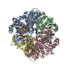
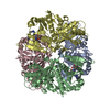
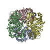
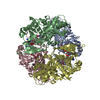
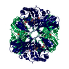
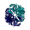
| Title | GLYCOSOMAL GLYCERALDEHYDE-3-PHOSPHATE DEHYDROGENASE IN A MONOCLINIC CRYSTAL FORM | ||||||
 Components Components | GLYCERALDEHYDE-3-PHOSPHATE DEHYDROGENASE | ||||||
 Keywords Keywords | OXIDOREDUCTASE / GLYCERALDEHYDE-3-PHOSPHATE DEHYDROGENASE / GLYCOSOME / TRYPANOSOME | ||||||
| Function / homology |  Function and homology information Function and homology informationglyceraldehyde-3-phosphate dehydrogenase (phosphorylating) / glycosome / glyceraldehyde-3-phosphate dehydrogenase (NAD+) (phosphorylating) activity / glycolytic process / glucose metabolic process / NAD binding / NADP binding / cytosol Similarity search - Function | ||||||
| Biological species |   Leishmania mexicana (eukaryote) Leishmania mexicana (eukaryote) | ||||||
| Method |  X-RAY DIFFRACTION / SYNCHROTRON / MOLECULAR REPLACEMENT / Resolution: 2.8 Å X-RAY DIFFRACTION / SYNCHROTRON / MOLECULAR REPLACEMENT / Resolution: 2.8 Å | ||||||
 Authors Authors | Kim, H. / Hol, W.G.J. | ||||||
 Citation Citation | Journal: J.Mol.Biol. / Year: 1998 Title: Crystal structure of Leishmania mexicana glycosomal glyceraldehyde-3-phosphate dehydrogenase in a new crystal form confirms the putative physiological active site structure. Authors: Kim, H. / Hol, W.G. #1: Journal: Biochemistry / Year: 1995Title: Crystal Structure of Glycosomal Glyceraldehyde-3-Phosphate Dehydrogenase from Leishmania Mexicana: Implications for Structure-Based Drug Design and a New Position for the Inorganic Phosphate Binding Site Authors: Kim, H. / Feil, I.K. / Verlinde, C.L. / Petra, P.H. / Hol, W.G. | ||||||
| History |
|
- Structure visualization
| Structure viewer | Molecule: MolmilJmol/JSmol |
|---|
- Downloads & links
-Download
| PDBx/mmCIF format | 1a7k.cif.gz | 276.1 KB | Display | PDBx/mmCIF format |
|---|---|---|---|---|
| PDB format | pdb1a7k.ent.gz | 228.3 KB | Display | PDB format |
| PDBx/mmJSON format | 1a7k.json.gz | Tree view | PDBx/mmJSON format | |
| Others |  Other downloads Other downloads |
-Validation report
| Summary document | 1a7k_validation.pdf.gz | 687.5 KB | Display | wwPDB validaton report |
|---|---|---|---|---|
| Full document | 1a7k_full_validation.pdf.gz | 726.4 KB | Display | |
| Data in XML | 1a7k_validation.xml.gz | 34.9 KB | Display | |
| Data in CIF | 1a7k_validation.cif.gz | 49.4 KB | Display | |
| Arichive directory | https://data.pdbj.org/pub/pdb/validation_reports/a7/1a7kftp://data.pdbj.org/pub/pdb/validation_reports/a7/1a7k | HTTPS FTP |
-Related structure data
| Similar structure data |
|---|

-Links
 PDBj
PDBj
- Assembly
| Deposited unit | 
| ||||||||
|---|---|---|---|---|---|---|---|---|---|
| 1 |
| ||||||||
| Unit cell |
| ||||||||
| Noncrystallographic symmetry (NCS) | NCS oper: (Code: given Matrix: (-0.999472, 0.030711, -0.010656), Vector: |
-Components
| #1: Protein | Mass: 38953.504 Da / Num. of mol.: 4 Source method: isolated from a genetically manipulated source Details: ONE BOUND NAD AND TWO BOUND PHOSPHATES PER MONOMER / Source: (gene. exp.) Leishmania mexicana (eukaryote) / Cell line: BL21 / Plasmid: PET-3A / Production host:  References: UniProt: Q27890, glyceraldehyde-3-phosphate dehydrogenase (phosphorylating) #2: Chemical | ChemComp-PO4 /   Mass: 94.971 Da / Num. of mol.: 8 / Source method: obtained synthetically / Formula: PO4 Mass: 94.971 Da / Num. of mol.: 8 / Source method: obtained synthetically / Formula: PO4#3: Chemical | ChemComp-NAD /   Mass: 663.425 Da / Num. of mol.: 4 / Source method: obtained synthetically / Formula: C21H27N7O14P2 / Comment: NAD*YM Mass: 663.425 Da / Num. of mol.: 4 / Source method: obtained synthetically / Formula: C21H27N7O14P2 / Comment: NAD*YM |
|---|
-Experimental details
-Experiment
| Experiment | Method: X-RAY DIFFRACTION / Number of used crystals: 1 |
|---|
- Sample preparation
| Crystal | Density Matthews: 2.32 Å3/Da / Density % sol: 46.94 % | ||||||||||||||||||||||||||||||||||||||||||||||||||||||
|---|---|---|---|---|---|---|---|---|---|---|---|---|---|---|---|---|---|---|---|---|---|---|---|---|---|---|---|---|---|---|---|---|---|---|---|---|---|---|---|---|---|---|---|---|---|---|---|---|---|---|---|---|---|---|---|
| Crystal grow | Method: vapor diffusion / pH: 7 Details: VAPOR DIFFUSION: 100 MM SODIUM PHOSPHATE, 27.5% PEG-1000 PH 7.0 (PH ADJUSTED WITH 100 MM CITRIC ACID), vapor diffusion | ||||||||||||||||||||||||||||||||||||||||||||||||||||||
| Crystal grow | *PLUS Method: vapor diffusion, sitting drop | ||||||||||||||||||||||||||||||||||||||||||||||||||||||
| Components of the solutions | *PLUS
|
-Data collection
| Diffraction | Mean temperature: 298 K |
|---|---|
| Diffraction source | Source: SYNCHROTRON / Site: CHESS  / Beamline: F1 / Wavelength: 0.98 / Beamline: F1 / Wavelength: 0.98 |
| Detector | Type: FUJI / Detector: IMAGE PLATE / Date: Oct 2, 1995 |
| Radiation | Monochromatic (M) / Laue (L): M / Scattering type: x-ray |
| Radiation wavelength | Wavelength: 0.98 Å / Relative weight: 1 |
| Reflection | Resolution: 2.8→20 Å / Num. obs: 28710 / % possible obs: 81 % / Redundancy: 2.5 % / Biso Wilson estimate: 52 Å2 / Rsym value: 0.071 |
| Reflection shell | Resolution: 2.8→2.9 Å / Redundancy: 4 % / Mean I/σ(I) obs: 1.8 / Rsym value: 0.481 / % possible all: 81 |
| Reflection | *PLUS Num. measured all: 78266 / Rmerge(I) obs: 0.071 |
| Reflection shell | *PLUS % possible obs: 81 % / Rmerge(I) obs: 0.481 |
- Processing
| Software |
| ||||||||||||||||||||||||||||||||||||||||||||||||||||||||||||
|---|---|---|---|---|---|---|---|---|---|---|---|---|---|---|---|---|---|---|---|---|---|---|---|---|---|---|---|---|---|---|---|---|---|---|---|---|---|---|---|---|---|---|---|---|---|---|---|---|---|---|---|---|---|---|---|---|---|---|---|---|---|
| Refinement | Method to determine structure: MOLECULAR REPLACEMENT Starting model: TRYPANOSOMA BRUCEI GAPDH Resolution: 2.8→8 Å / Cross valid method: THROUGHOUT
| ||||||||||||||||||||||||||||||||||||||||||||||||||||||||||||
| Displacement parameters | Biso mean: 49 Å2 | ||||||||||||||||||||||||||||||||||||||||||||||||||||||||||||
| Refinement step | Cycle: LAST / Resolution: 2.8→8 Å
| ||||||||||||||||||||||||||||||||||||||||||||||||||||||||||||
| Refine LS restraints |
| ||||||||||||||||||||||||||||||||||||||||||||||||||||||||||||
| LS refinement shell | Highest resolution: 2.8 Å / Total num. of bins used: 8
| ||||||||||||||||||||||||||||||||||||||||||||||||||||||||||||
| Xplor file |
| ||||||||||||||||||||||||||||||||||||||||||||||||||||||||||||
| Software | *PLUS Name: X-PLOR / Version: 3 / Classification: refinement | ||||||||||||||||||||||||||||||||||||||||||||||||||||||||||||
| Refinement | *PLUS | ||||||||||||||||||||||||||||||||||||||||||||||||||||||||||||
| Solvent computation | *PLUS | ||||||||||||||||||||||||||||||||||||||||||||||||||||||||||||
| Displacement parameters | *PLUS | ||||||||||||||||||||||||||||||||||||||||||||||||||||||||||||
| Refine LS restraints | *PLUS
| ||||||||||||||||||||||||||||||||||||||||||||||||||||||||||||
| LS refinement shell | *PLUS Rfactor obs: 0.338 |
