 Movie
Movie Controller

[English] 日本語
 Yorodumi
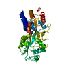
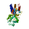
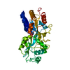
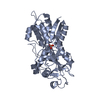
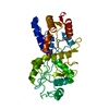
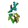
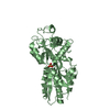
- PDB-1a54: PHOSPHATE-BINDING PROTEIN MUTANT A197C LABELLED WITH A COUMARIN F... -
+ Open data

- Basic information
| Entry | Database: PDB / ID: 1a54 | |||||||||
|---|---|---|---|---|---|---|---|---|---|---|
| Title | PHOSPHATE-BINDING PROTEIN MUTANT A197C LABELLED WITH A COUMARIN FLUOROPHORE AND BOUND TO DIHYDROGENPHOSPHATE ION | |||||||||
 Components Components | Phosphate-binding protein PstS | |||||||||
 Keywords Keywords | PHOSPHOTRANSFERASE / TRANSPORT / COUMARIN / FLUOROPHORE | |||||||||
| Function / homology |  Function and homology information Function and homology informationphosphate ion transport / phosphate ion transmembrane transport / phosphate ion binding / ATP-binding cassette (ABC) transporter complex, substrate-binding subunit-containing / ATP-binding cassette (ABC) transporter complex / response to radiation / outer membrane-bounded periplasmic space / DNA damage response / membrane Similarity search - Function | |||||||||
| Biological species |  | |||||||||
| Method |  X-RAY DIFFRACTION / SYNCHROTRON / molecular replacement / Resolution: 1.6 Å X-RAY DIFFRACTION / SYNCHROTRON / molecular replacement / Resolution: 1.6 Å | |||||||||
 Authors Authors | Hirshberg, M. / Henrick, K. / Lloyd-Haire, L. / Vasisht, N. / Brune, M. / Corrie, J.E.T. / Webb, M.R. | |||||||||
 Citation Citation | Journal: Biochemistry / Year: 1998 Title: Crystal structure of phosphate binding protein labeled with a coumarin fluorophore, a probe for inorganic phosphate. Authors: Hirshberg, M. / Henrick, K. / Haire, L.L. / Vasisht, N. / Brune, M. / Corrie, J.E. / Webb, M.R. #1: Journal: Biochemistry / Year: 1998Title: Mechanism of Inorganic Phosphate Interaction with Phosphate Binding Protein from Escherichia Coli Authors: Brune, M. / Hunter, J.L. / Howell, S.A. / Martin, S.R. / Hazlett, T.L. / Corrie, J.E. / Webb, M.R. | |||||||||
| History |
|
- Structure visualization
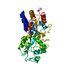
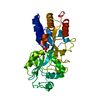
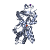
| Structure viewer | Molecule: MolmilJmol/JSmol |
|---|
- Downloads & links
-Download
| PDBx/mmCIF format | 1a54.cif.gz | 81.3 KB | Display | PDBx/mmCIF format |
|---|---|---|---|---|
| PDB format | pdb1a54.ent.gz | 60.5 KB | Display | PDB format |
| PDBx/mmJSON format | 1a54.json.gz | Tree view | PDBx/mmJSON format | |
| Others |  Other downloads Other downloads |
-Validation report
| Arichive directory | https://data.pdbj.org/pub/pdb/validation_reports/a5/1a54ftp://data.pdbj.org/pub/pdb/validation_reports/a5/1a54 | HTTPS FTP |
|---|
-Related structure data
| Related structure data |  1a55C 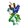 2abhS S: Starting model for refinement C: citing same article ( |
|---|---|
| Similar structure data |
-Links
 PDBj
PDBj- Assembly
| Deposited unit | 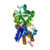
| ||||||||
|---|---|---|---|---|---|---|---|---|---|
| 1 |
| ||||||||
| Unit cell |
|
-Components
| #1: Protein | Mass: 34489.664 Da / Num. of mol.: 1 / Mutation: A197C Source method: isolated from a genetically manipulated source Source: (gene. exp.) |
|---|---|
| #2: Chemical | ChemComp-2HP /   Mass: 96.987 Da / Num. of mol.: 1 / Source method: obtained synthetically / Formula: H2O4P Mass: 96.987 Da / Num. of mol.: 1 / Source method: obtained synthetically / Formula: H2O4P |
| #3: Chemical | ChemComp-MDC /   Mass: 383.398 Da / Num. of mol.: 1 / Source method: obtained synthetically / Formula: C20H21N3O5 Mass: 383.398 Da / Num. of mol.: 1 / Source method: obtained synthetically / Formula: C20H21N3O5 |
| #4: Water | ChemComp-HOH /  Mass: 18.015 Da / Num. of mol.: 334 / Source method: isolated from a natural source / Formula: H2O Mass: 18.015 Da / Num. of mol.: 334 / Source method: isolated from a natural source / Formula: H2O |
| Has protein modification | N |
-Experimental details
-Experiment
| Experiment | Method: X-RAY DIFFRACTION / Number of used crystals: 1 |
|---|
- Sample preparation
| Crystal | Density Matthews: 2.31 Å3/Da / Density % sol: 46.74 % | ||||||||||||||||||||||||||||||||||||||||||
|---|---|---|---|---|---|---|---|---|---|---|---|---|---|---|---|---|---|---|---|---|---|---|---|---|---|---|---|---|---|---|---|---|---|---|---|---|---|---|---|---|---|---|---|
| Crystal grow | pH: 4.5 / Details: pH 4.5 | ||||||||||||||||||||||||||||||||||||||||||
| Crystal grow | *PLUS pH: 7.6 / Method: vapor diffusion, hanging drop / Details: used to seeding | ||||||||||||||||||||||||||||||||||||||||||
| Components of the solutions | *PLUS
|
-Data collection
| Diffraction | Mean temperature: 102 K |
|---|---|
| Diffraction source | Source: SYNCHROTRON / Site: SRS  / Beamline: PX9.5 / Wavelength: 0.8 / Beamline: PX9.5 / Wavelength: 0.8 |
| Detector | Type: MARRESEARCH / Detector: IMAGE PLATE / Date: Aug 1, 1997 |
| Radiation | Monochromatic (M) / Laue (L): M / Scattering type: x-ray |
| Radiation wavelength | Wavelength: 0.8 Å / Relative weight: 1 |
| Reflection | Resolution: 1.6→12 Å / Num. obs: 345731 / % possible obs: 95.5 % / Observed criterion σ(I): 0 / Redundancy: 3.3 % / Rmerge(I) obs: 0.08 |
| Reflection shell | Highest resolution: 1.6 Å / Redundancy: 2.9 % / Rmerge(I) obs: 0.21 / % possible all: 93.4 |
| Reflection | *PLUS Num. obs: 40945 / Num. measured all: 345731 |
| Reflection shell | *PLUS % possible obs: 93.4 % / Mean I/σ(I) obs: 3.8 |
- Processing
| Software |
| |||||||||||||||||||||||||||||||||||||||||||||||||||||||||||||||
|---|---|---|---|---|---|---|---|---|---|---|---|---|---|---|---|---|---|---|---|---|---|---|---|---|---|---|---|---|---|---|---|---|---|---|---|---|---|---|---|---|---|---|---|---|---|---|---|---|---|---|---|---|---|---|---|---|---|---|---|---|---|---|---|---|
| Refinement | Method to determine structure: molecular replacement Starting model: PDB ENTRY 2ABH Resolution: 1.6→12 Å / Cross valid method: THROUGHOUT
| |||||||||||||||||||||||||||||||||||||||||||||||||||||||||||||||
| Refinement step | Cycle: LAST / Resolution: 1.6→12 Å
| |||||||||||||||||||||||||||||||||||||||||||||||||||||||||||||||
| Refine LS restraints |
| |||||||||||||||||||||||||||||||||||||||||||||||||||||||||||||||
| Software | *PLUS Name: REFMAC / Classification: refinement | |||||||||||||||||||||||||||||||||||||||||||||||||||||||||||||||
| Refinement | *PLUS Rfactor obs: 0.177 | |||||||||||||||||||||||||||||||||||||||||||||||||||||||||||||||
| Solvent computation | *PLUS | |||||||||||||||||||||||||||||||||||||||||||||||||||||||||||||||
| Displacement parameters | *PLUS Biso mean: 14.9 Å2 |
