 Movie
Movie Controller


+ Open data

- Basic information
| Entry | Database: PDB / ID: 1a3z | ||||||
|---|---|---|---|---|---|---|---|
| Title | REDUCED RUSTICYANIN AT 1.9 ANGSTROMS | ||||||
 Components Components | RUSTICYANIN | ||||||
 Keywords Keywords | ELECTRON TRANSPORT / CUPREDOXIN / METALLOPROTEIN / REDOX POTENTIAL / ACIDOPHILIC | ||||||
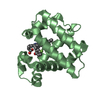
| Function / homology |  Function and homology information Function and homology information | ||||||
| Biological species |  Acidithiobacillus ferrooxidans (bacteria) Acidithiobacillus ferrooxidans (bacteria) | ||||||
| Method |  X-RAY DIFFRACTION / PHASES TAKEN FROM THE OXIDIZED FORM, PDB ENTRY 1RCY / Resolution: 1.9 Å X-RAY DIFFRACTION / PHASES TAKEN FROM THE OXIDIZED FORM, PDB ENTRY 1RCY / Resolution: 1.9 Å | ||||||
 Authors Authors | Zhao, D. / Shoham, M. | ||||||
 Citation Citation | Journal: Biophys.J. / Year: 1998 Title: Rusticyanin: Extremes in acid stability and redox potential explained by the crystal structure. Authors: Zhao, D. / Shoham, M. #1: Journal: J.Mol.Biol. / Year: 1996Title: Multiple Wavelength Anomalous Diffraction (MAD) Crystal Structure of Rusticyanin: A Highly Oxidizing Cupredoxin with Extreme Acid Stability Authors: Walter, R.L. / Ealick, S.E. / Friedman, A.M. / Blake II, R.C. / Proctor, P. / Shoham, M. #2: Journal: J.Mol.Biol. / Year: 1996Title: NMR Solution Structure of Cu(I) Rusticyanin from Thiobacillus Ferrooxidans: Structural Basis for the Extreme Acid Stability and Redox Potential Authors: Botuyan, M.V. / Toy-Palmer, A. / Chung, J. / Blake II, R.C. / Beroza, P. / Case, D.A. / Dyson, H.J. #3: Journal: J.Mol.Biol. / Year: 1992Title: Crystallization and Preliminary X-Ray Crystallographic Studies of Rusticyanin from Thiobacillus Ferrooxidans Authors: Djebli, A. / Proctor, P. / Blake II, R.C. / Shoham, M. | ||||||
| History |
|
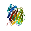
- Structure visualization
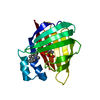
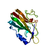
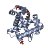
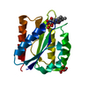
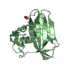
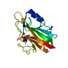
| Structure viewer | Molecule: MolmilJmol/JSmol |
|---|
- Downloads & links
-Download
| PDBx/mmCIF format | 1a3z.cif.gz | 42.7 KB | Display | PDBx/mmCIF format |
|---|---|---|---|---|
| PDB format | pdb1a3z.ent.gz | 29.4 KB | Display | PDB format |
| PDBx/mmJSON format | 1a3z.json.gz | Tree view | PDBx/mmJSON format | |
| Others |  Other downloads Other downloads |
-Validation report
| Summary document | 1a3z_validation.pdf.gz | 419.9 KB | Display | wwPDB validaton report |
|---|---|---|---|---|
| Full document | 1a3z_full_validation.pdf.gz | 421.3 KB | Display | |
| Data in XML | 1a3z_validation.xml.gz | 8.7 KB | Display | |
| Data in CIF | 1a3z_validation.cif.gz | 11.3 KB | Display | |
| Arichive directory | https://data.pdbj.org/pub/pdb/validation_reports/a3/1a3zftp://data.pdbj.org/pub/pdb/validation_reports/a3/1a3z | HTTPS FTP |
-Related structure data
| Related structure data |  1rcyS S: Starting model for refinement |
|---|---|
| Similar structure data |

-Links
 PDBj
PDBj- Assembly
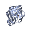
| Deposited unit | 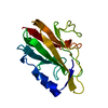
| ||||||||
|---|---|---|---|---|---|---|---|---|---|
| 1 |
| ||||||||
| Unit cell |
|
-Components
| #1: Protein | Mass: 16569.879 Da / Num. of mol.: 1 / Source method: isolated from a natural source / Details: REDUCED FORM / Source: (natural) Acidithiobacillus ferrooxidans (bacteria) / Cellular location: PERIPLASM / References: UniProt: P24930, UniProt: P0C918*PLUS |
|---|---|
| #2: Chemical | ChemComp-CU1 /   Mass: 63.546 Da / Num. of mol.: 1 / Source method: obtained synthetically / Formula: Cu Mass: 63.546 Da / Num. of mol.: 1 / Source method: obtained synthetically / Formula: Cu |
| #3: Water | ChemComp-HOH /  Mass: 18.015 Da / Num. of mol.: 85 / Source method: isolated from a natural source / Formula: H2O Mass: 18.015 Da / Num. of mol.: 85 / Source method: isolated from a natural source / Formula: H2O |
-Experimental details
-Experiment
| Experiment | Method: X-RAY DIFFRACTION / Number of used crystals: 1 |
|---|
- Sample preparation
| Crystal | Density Matthews: 2.22 Å3/Da / Density % sol: 44 % |
|---|---|
| Crystal grow | Method: vapor diffusion, hanging drop / pH: 4.6 Details: VAPOR DIFFUSION IN HANGING DROPS AGAINST 100 MM-SODIUM CITRATE, PH 4.6, 200 MM LITHIUM CHLORIDE AND 25%(W/V) PEG 8000 AT 277K. CRYSTALS SOAKED IN 10MM DITHIONITE FOR 3 DAYS PRIOR TO DATA ...Details: VAPOR DIFFUSION IN HANGING DROPS AGAINST 100 MM-SODIUM CITRATE, PH 4.6, 200 MM LITHIUM CHLORIDE AND 25%(W/V) PEG 8000 AT 277K. CRYSTALS SOAKED IN 10MM DITHIONITE FOR 3 DAYS PRIOR TO DATA COLLECTION., vapor diffusion - hanging drop |
-Data collection
| Diffraction | Mean temperature: 293 K |
|---|---|
| Diffraction source | Source: ROTATING ANODE / Type: RIGAKU RUH2R / Wavelength: 1.5418 |
| Detector | Type: ADSC / Detector: AREA DETECTOR / Date: Jun 1, 1995 |
| Radiation | Monochromator: GRAPHITE(002) / Monochromatic (M) / Laue (L): M / Scattering type: x-ray |
| Radiation wavelength | Wavelength: 1.5418 Å / Relative weight: 1 |
| Reflection | Resolution: 1.9→100 Å / Num. obs: 9718 / % possible obs: 87.4 % / Observed criterion σ(I): 0 / Redundancy: 1.97 % / Rmerge(I) obs: 0.063 / Rsym value: 0.063 / Net I/σ(I): 13.3 |
| Reflection shell | Resolution: 1.9→1.969 Å / Redundancy: 1.42 % / Rmerge(I) obs: 0.199 / Mean I/σ(I) obs: 3.02 / Rsym value: 0.199 / % possible all: 78 |
- Processing
| Software |
| ||||||||||||||||||||||||||||||||||||||||||||||||||||||||||||
|---|---|---|---|---|---|---|---|---|---|---|---|---|---|---|---|---|---|---|---|---|---|---|---|---|---|---|---|---|---|---|---|---|---|---|---|---|---|---|---|---|---|---|---|---|---|---|---|---|---|---|---|---|---|---|---|---|---|---|---|---|---|
| Refinement | Method to determine structure: PHASES TAKEN FROM THE OXIDIZED FORM, PDB ENTRY 1RCY Starting model: PDB ENTRY 1RCY Resolution: 1.9→100 Å / Data cutoff high absF: 1000000 / Data cutoff low absF: 0.001 / Isotropic thermal model: RESTRAINED / Cross valid method: THROUGHOUT / σ(F): 0
| ||||||||||||||||||||||||||||||||||||||||||||||||||||||||||||
| Displacement parameters | Biso mean: 19 Å2 | ||||||||||||||||||||||||||||||||||||||||||||||||||||||||||||
| Refinement step | Cycle: LAST / Resolution: 1.9→100 Å
| ||||||||||||||||||||||||||||||||||||||||||||||||||||||||||||
| Refine LS restraints |
| ||||||||||||||||||||||||||||||||||||||||||||||||||||||||||||
| LS refinement shell | Resolution: 1.9→1.99 Å / Total num. of bins used: 8
| ||||||||||||||||||||||||||||||||||||||||||||||||||||||||||||
| Xplor file |
|
