 Movie
Movie Controller

[English] 日本語
 Yorodumi
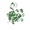
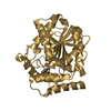
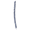
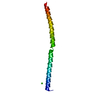
- PDB-1a3i: X-RAY CRYSTALLOGRAPHIC DETERMINATION OF A COLLAGEN-LIKE PEPTIDE W... -
+ Open data

- Basic information
| Entry | Database: PDB / ID: 1a3i | ||||||
|---|---|---|---|---|---|---|---|
| Title | X-RAY CRYSTALLOGRAPHIC DETERMINATION OF A COLLAGEN-LIKE PEPTIDE WITH THE REPEATING SEQUENCE (PRO-PRO-GLY) | ||||||
 Components Components | (COLLAGEN-LIKE PEPTIDE) x 2 | ||||||
 Keywords Keywords | EXTRACELLULAR MATRIX / COLLAGEN | ||||||
| Function / homology | ACETIC ACID Function and homology information Function and homology information | ||||||
| Method |  X-RAY DIFFRACTION / MOLECULAR REPLACEMENT / Resolution: 1.97 Å X-RAY DIFFRACTION / MOLECULAR REPLACEMENT / Resolution: 1.97 Å | ||||||
 Authors Authors | Kramer, R.Z. / Vitagliano, L. / Bella, J. / Berisio, R. / Mazzarella, L. / Brodsky, B. / Zagari, A. / Berman, H.M. | ||||||
 Citation Citation | Journal: J.Mol.Biol. / Year: 1998 Title: X-ray crystallographic determination of a collagen-like peptide with the repeating sequence (Pro-Pro-Gly). Authors: Kramer, R.Z. / Vitagliano, L. / Bella, J. / Berisio, R. / Mazzarella, L. / Brodsky, B. / Zagari, A. / Berman, H.M. | ||||||
| History |
|
- Structure visualization
| Structure viewer | Molecule: MolmilJmol/JSmol |
|---|
- Downloads & links
-Download
| PDBx/mmCIF format | 1a3i.cif.gz | 11.8 KB | Display | PDBx/mmCIF format |
|---|---|---|---|---|
| PDB format | pdb1a3i.ent.gz | 8 KB | Display | PDB format |
| PDBx/mmJSON format | 1a3i.json.gz | Tree view | PDBx/mmJSON format | |
| Others |  Other downloads Other downloads |
-Validation report
| Arichive directory | https://data.pdbj.org/pub/pdb/validation_reports/a3/1a3iftp://data.pdbj.org/pub/pdb/validation_reports/a3/1a3i | HTTPS FTP |
|---|
-Related structure data


-Links
 PDBj
PDBj

- Assembly
| Deposited unit | 
| ||||||||
|---|---|---|---|---|---|---|---|---|---|
| 1 | x 5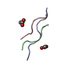
| ||||||||
| Unit cell |
| ||||||||
| Details | THE 21 RESIDUE ASYMMETRIC UNIT CORRESPONDS TO ONE TRIPLE-HELICAL REPEAT AND IS SMALLER THAN THE ENTIRE 90 RESIDUE PEPTIDE DUE TO TRANSLATIONAL DISORDER ALONG THE HELICAL AXIS. THE RESULT IS A POLYMER-LIKE STRUCTURE WITH NO DEFINED ENDS. THE POLYMER STRUCTURE IS FORMED BY CONTINUATION OF THE CHAINS USING THE SYMMETRY-RELATED MOLECULES ALONG THE HELICAL AXIS. THE TVECT RECORD BELOW PRESENTS THE TRANSLATION THAT WILL GENERATE THE POLYMER. NOTE: THEREFORE, CLOSE CONTACTS BETWEEN SYMMETRY-RELATED MOLECULES ARE INTENTIONAL AND NECESSARY. INTERCHAIN HYDROGEN BONDING AT THE END OF CHAINS ALSO UTILIZES SYMMETRY-RELATED MOLECULES. THE ENTIRE 30 RESIDUE LONG PEPTIDE CAN BE GENERATED FROM THE SUBMITTED ASYMMETRIC UNIT BY APPLYING THE FOLLOWING TRANSLATIONS (USING FRACTIONAL COORDINATES): CHAIN A: TRANSLATE RESIDUES 1 - 9 BY (0 0 1), (0 0 2), AND (0 0 3) AND RESIDUES 7 - 9 BY (0 0 4). CHAIN B: TRANSLATE RESIDUES 31 - 36 BY (0 0 1), (0 0 2), AND (0 0 3). CHAIN C: TRANSLATE RESIDUES 61 - 66 BY (0 0 1), (0 0 2), AND (0 0 3) AND RESIDUES 64 - 66 BY (004). THIS WILL RESULT IN A MOLECULE WITH A TOTAL OF 90 RESIDUES, 30 IN EACH CHAIN. |
-Components
| #1: Protein/peptide | Mass: 771.859 Da / Num. of mol.: 1 Source method: isolated from a genetically manipulated source | ||||||||||
|---|---|---|---|---|---|---|---|---|---|---|---|
| #2: Protein/peptide | Mass: 520.578 Da / Num. of mol.: 2 Source method: isolated from a genetically manipulated source #3: Chemical |   Mass: 60.052 Da / Num. of mol.: 2 / Source method: obtained synthetically / Formula: C2H4O2 Mass: 60.052 Da / Num. of mol.: 2 / Source method: obtained synthetically / Formula: C2H4O2#4: Water | ChemComp-HOH / |  Mass: 18.015 Da / Num. of mol.: 37 / Source method: isolated from a natural source / Formula: H2O Mass: 18.015 Da / Num. of mol.: 37 / Source method: isolated from a natural source / Formula: H2OCompound details | HYDROGEN BONDS BETWEEN PEPTIDE CHAINS FOLLOW THE RICH AND CRICK MODEL II FOR COLLAGEN. | Has protein modification | Y | Sequence details | FOR EACH CHAIN, RESIDUE NUMBERING CORRESPOND | |
-Experimental details
-Experiment
| Experiment | Method: X-RAY DIFFRACTION / Number of used crystals: 1 |
|---|
- Sample preparation
| Crystal | Density Matthews: 1.95 Å3/Da / Density % sol: 36.82 % | ||||||||||||||||||||||||||||||
|---|---|---|---|---|---|---|---|---|---|---|---|---|---|---|---|---|---|---|---|---|---|---|---|---|---|---|---|---|---|---|---|
| Crystal grow | Details: PEPTIDE WAS CRYSTALLIZED FROM 4.0 MG/ML PEPTIDE IN 10% ACETIC ACID, 0.1% SODIUM AZIDE, AND 3.0% PEG400. | ||||||||||||||||||||||||||||||
| Crystal grow | *PLUS Temperature: 4 ℃ / Method: vapor diffusion, hanging drop | ||||||||||||||||||||||||||||||
| Components of the solutions | *PLUS
|
-Data collection
| Diffraction | Mean temperature: 259 K |
|---|---|
| Diffraction source | Wavelength: 1.5418 |
| Detector | Type: ENRAF-NONIUS FAST / Detector: DIFFRACTOMETER / Date: Oct 1, 1991 |
| Radiation | Monochromatic (M) / Laue (L): M / Scattering type: x-ray |
| Radiation wavelength | Wavelength: 1.5418 Å / Relative weight: 1 |
| Reflection | Highest resolution: 1.97 Å / Num. obs: 1136 / % possible obs: 99.5 % / Observed criterion σ(I): 0 |
| Reflection shell | Resolution: 1.97→2.2 Å / % possible all: 98 |
| Reflection | *PLUS % possible obs: 100 % |
| Reflection shell | *PLUS Rmerge(I) obs: 98 |
- Processing
| Software |
| ||||||||||||||||||||||||||||||||||||||||||||||||||||||||||||
|---|---|---|---|---|---|---|---|---|---|---|---|---|---|---|---|---|---|---|---|---|---|---|---|---|---|---|---|---|---|---|---|---|---|---|---|---|---|---|---|---|---|---|---|---|---|---|---|---|---|---|---|---|---|---|---|---|---|---|---|---|---|
| Refinement | Method to determine structure: MOLECULAR REPLACEMENT Starting model: IDEALIZED SEVEN-FOLD TRIPLE-HELIX Resolution: 1.97→8 Å / Data cutoff high absF: 1000000 / Data cutoff low absF: 0.1 / Isotropic thermal model: GROUP / σ(F): 2 Details: DUE TO THE QUASI-INFINITE, AVERAGED NATURE OF THE TRIPLE HELIX, DURING REFINEMENT COVALENT BONDS ARE NECESSARY TO JOIN THE MOLECULE WITH ITS SYMMETRY MATES BOTH ABOVE IT AND BELOW IT ALONG ...Details: DUE TO THE QUASI-INFINITE, AVERAGED NATURE OF THE TRIPLE HELIX, DURING REFINEMENT COVALENT BONDS ARE NECESSARY TO JOIN THE MOLECULE WITH ITS SYMMETRY MATES BOTH ABOVE IT AND BELOW IT ALONG THE HELICAL AXIS AND TIGHT REFINEMENT CONSTRAINTS WERE MAINTAINED. THE UNIT CELL AXES WERE CHOSEN TO COINCIDE WITH A PREVIOUS STRUCTURE DETERMINATION (OKUYAMA 1981) OF THIS PEPTIDE.
| ||||||||||||||||||||||||||||||||||||||||||||||||||||||||||||
| Displacement parameters | Biso mean: 15.9 Å2 | ||||||||||||||||||||||||||||||||||||||||||||||||||||||||||||
| Refinement step | Cycle: LAST / Resolution: 1.97→8 Å
| ||||||||||||||||||||||||||||||||||||||||||||||||||||||||||||
| Refine LS restraints |
| ||||||||||||||||||||||||||||||||||||||||||||||||||||||||||||
| LS refinement shell | Resolution: 1.97→2.04 Å / Total num. of bins used: 10
| ||||||||||||||||||||||||||||||||||||||||||||||||||||||||||||
| Xplor file |
| ||||||||||||||||||||||||||||||||||||||||||||||||||||||||||||
| Software | *PLUS Name: X-PLOR / Version: 3.1 / Classification: refinement | ||||||||||||||||||||||||||||||||||||||||||||||||||||||||||||
| Refine LS restraints | *PLUS
|
