 Movie
Movie Controller


+ Open data

- Basic information
| Entry | Database: PDB / ID: 1a3f | ||||||
|---|---|---|---|---|---|---|---|
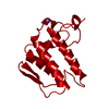
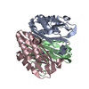
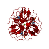
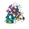
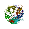
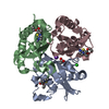
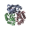
| Title | PHOSPHOLIPASE A2 (PLA2) FROM NAJA NAJA VENOM | ||||||
 Components Components | PHOSPHOLIPASE A2 | ||||||
 Keywords Keywords | CARBOXYLIC ESTER HYDROLASE / PHOSPHOLIPASE / TRIMER / CALCIUM BINDING / ACTIVATOR SITE | ||||||
| Function / homology |  Function and homology information Function and homology information: / phospholipase A2 / arachidonate secretion / lipid catabolic process / phospholipid metabolic process / phospholipid binding / calcium ion binding / extracellular region Similarity search - Function | ||||||
| Biological species |  Naja naja (Indian cobra) Naja naja (Indian cobra) | ||||||
| Method |  X-RAY DIFFRACTION / MOLECULAR REPLACEMENT / Resolution: 2.65 Å X-RAY DIFFRACTION / MOLECULAR REPLACEMENT / Resolution: 2.65 Å | ||||||
 Authors Authors | Segelke, B.W. / Nguyen, D. / Chee, R. / Xuong, H.N. / Dennis, E.A. | ||||||
 Citation Citation | Journal: J.Mol.Biol. / Year: 1998 Title: Structures of two novel crystal forms of Naja naja naja phospholipase A2 lacking Ca2+ reveal trimeric packing. Authors: Segelke, B.W. / Nguyen, D. / Chee, R. / Xuong, N.H. / Dennis, E.A. | ||||||
| History |
|
- Structure visualization
| Structure viewer | Molecule: MolmilJmol/JSmol |
|---|
- Downloads & links
-Download
| PDBx/mmCIF format | 1a3f.cif.gz | 76.9 KB | Display | PDBx/mmCIF format |
|---|---|---|---|---|
| PDB format | pdb1a3f.ent.gz | 59.6 KB | Display | PDB format |
| PDBx/mmJSON format | 1a3f.json.gz | Tree view | PDBx/mmJSON format | |
| Others |  Other downloads Other downloads |
-Validation report
| Arichive directory | https://data.pdbj.org/pub/pdb/validation_reports/a3/1a3fftp://data.pdbj.org/pub/pdb/validation_reports/a3/1a3f | HTTPS FTP |
|---|
-Related structure data
| Related structure data |  1a3dC 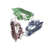 1pshS S: Starting model for refinement C: citing same article ( |
|---|---|
| Similar structure data |


-Links
 PDBj
PDBj

- Assembly
| Deposited unit | 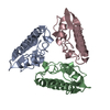
| ||||||||||||||||||||||||
|---|---|---|---|---|---|---|---|---|---|---|---|---|---|---|---|---|---|---|---|---|---|---|---|---|---|
| 1 |
| ||||||||||||||||||||||||
| Unit cell |
| ||||||||||||||||||||||||
| Noncrystallographic symmetry (NCS) | NCS domain:
NCS oper:
|
-Components
| #1: Protein | Mass: 13357.855 Da / Num. of mol.: 3 / Source method: isolated from a natural source / Source: (natural) Naja naja (Indian cobra) / Cellular location: VENOM SACK / References: UniProt: P15445, phospholipase A2Has protein modification | Y | |
|---|
-Experimental details
-Experiment
| Experiment | Method: X-RAY DIFFRACTION / Number of used crystals: 1 |
|---|
- Sample preparation
| Crystal | Density Matthews: 2.67 Å3/Da / Density % sol: 53.4 % | |||||||||||||||
|---|---|---|---|---|---|---|---|---|---|---|---|---|---|---|---|---|
| Crystal grow | Temperature: 295.5 K Details: UNBUFFERED 32.5% MONOMETHYL PEG 5K AND 0.17M SODIUM CITRATE AT 22.5 DEGREES C., temperature 295.5K | |||||||||||||||
| Crystal | *PLUS Density % sol: 54 % | |||||||||||||||
| Crystal grow | *PLUS Temperature: 22.5 ℃ / Method: vapor diffusion, hanging drop | |||||||||||||||
| Components of the solutions | *PLUS
|
-Data collection
| Diffraction | Mean temperature: 287 K |
|---|---|
| Diffraction source | Source: ROTATING ANODE / Type: RIGAKU / Wavelength: 1.5418 |
| Detector | Type: XUONG-HAMLIN MULTIWIRE / Detector: AREA DETECTOR / Date: Jun 1, 1995 |
| Radiation | Monochromator: GRAPHITE(002) / Monochromatic (M) / Laue (L): M / Scattering type: x-ray |
| Radiation wavelength | Wavelength: 1.5418 Å / Relative weight: 1 |
| Reflection | Resolution: 2.5→72.5 Å / Num. obs: 12166 / % possible obs: 79 % / Observed criterion σ(I): 2 / Redundancy: 2.5 % / Biso Wilson estimate: 14.7 Å2 / Rsym value: 0.064 / Net I/σ(I): 8.7 |
| Reflection shell | Resolution: 2.5→2.69 Å / Redundancy: 1.4 % / Mean I/σ(I) obs: 2 / Rsym value: 0.24 / % possible all: 73 |
| Reflection | *PLUS Num. obs: 9506 / % possible obs: 72.4 % / Num. measured all: 27711 / Rmerge(I) obs: 0.064 |
- Processing
| Software |
| ||||||||||||||||||||||||||||||||||||||||||||||||||||||||||||
|---|---|---|---|---|---|---|---|---|---|---|---|---|---|---|---|---|---|---|---|---|---|---|---|---|---|---|---|---|---|---|---|---|---|---|---|---|---|---|---|---|---|---|---|---|---|---|---|---|---|---|---|---|---|---|---|---|---|---|---|---|---|
| Refinement | Method to determine structure: MOLECULAR REPLACEMENT Starting model: PDB ENTRY 1PSH Resolution: 2.65→8 Å / Rfactor Rfree error: 0.008 / Data cutoff high absF: 1000000 / Data cutoff low absF: 0.001 / Isotropic thermal model: RESTRAINED / Cross valid method: POST REFINEMENT / σ(F): 2
| ||||||||||||||||||||||||||||||||||||||||||||||||||||||||||||
| Displacement parameters | Biso mean: 16.3 Å2 | ||||||||||||||||||||||||||||||||||||||||||||||||||||||||||||
| Refine analyze | Luzzati coordinate error obs: 0.3 Å / Luzzati d res low obs: 8 Å / Luzzati sigma a obs: 0.33 Å | ||||||||||||||||||||||||||||||||||||||||||||||||||||||||||||
| Refinement step | Cycle: LAST / Resolution: 2.65→8 Å
| ||||||||||||||||||||||||||||||||||||||||||||||||||||||||||||
| Refine LS restraints |
| ||||||||||||||||||||||||||||||||||||||||||||||||||||||||||||
| Refine LS restraints NCS | Refine-ID: X-RAY DIFFRACTION / Weight position: 250
| ||||||||||||||||||||||||||||||||||||||||||||||||||||||||||||
| LS refinement shell | Resolution: 2.65→2.77 Å / Rfactor Rfree error: 0.036 / Total num. of bins used: 8
| ||||||||||||||||||||||||||||||||||||||||||||||||||||||||||||
| Xplor file |
| ||||||||||||||||||||||||||||||||||||||||||||||||||||||||||||
| Software | *PLUS Name: X-PLOR / Version: 3 / Classification: refinement | ||||||||||||||||||||||||||||||||||||||||||||||||||||||||||||
| Refine LS restraints | *PLUS
|
