 Movie
Movie Controller

[English] 日本語
 Yorodumi
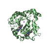
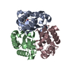
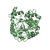
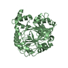
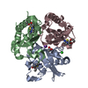
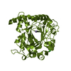
- PDB-1f7r: CRYSTAL STRUCTURES OF FELINE IMMUNODEFICIENCY VIRUS DUTP PYROPHOS... -
+ Open data

- Basic information
| Entry | Database: PDB / ID: 1f7r | ||||||
|---|---|---|---|---|---|---|---|
| Title | CRYSTAL STRUCTURES OF FELINE IMMUNODEFICIENCY VIRUS DUTP PYROPHOSPHATASE AND ITS NUCLEOTIDE COMPLEXES IN THREE CRYSTAL FORMS. | ||||||
 Components Components | POL POLYPROTEIN | ||||||
 Keywords Keywords | Viral protein / hydrolase / Eight stranded beta barrel protein | ||||||
| Function / homology |  Function and homology information Function and homology informationdUTP catabolic process / dUMP biosynthetic process / dUTP diphosphatase / dUTP diphosphatase activity / Hydrolases; Acting on peptide bonds (peptidases); Aspartic endopeptidases / retroviral ribonuclease H / exoribonuclease H / exoribonuclease H activity / DNA integration / viral genome integration into host DNA ...dUTP catabolic process / dUMP biosynthetic process / dUTP diphosphatase / dUTP diphosphatase activity / Hydrolases; Acting on peptide bonds (peptidases); Aspartic endopeptidases / retroviral ribonuclease H / exoribonuclease H / exoribonuclease H activity / DNA integration / viral genome integration into host DNA / establishment of integrated proviral latency / RNA-directed DNA polymerase / RNA stem-loop binding / RNA-directed DNA polymerase activity / RNA-DNA hybrid ribonuclease activity / Transferases; Transferring phosphorus-containing groups; Nucleotidyltransferases / DNA recombination / aspartic-type endopeptidase activity / Hydrolases; Acting on ester bonds / symbiont entry into host cell / magnesium ion binding / proteolysis / DNA binding / zinc ion binding Similarity search - Function | ||||||
| Biological species |  Feline immunodeficiency virus Feline immunodeficiency virus | ||||||
| Method |  X-RAY DIFFRACTION / Resolution: 2.5 Å X-RAY DIFFRACTION / Resolution: 2.5 Å | ||||||
 Authors Authors | Prasad, G.S. / Stura, E.A. / Elder, J.H. / Stout, C.D. | ||||||
 Citation Citation | Journal: Acta Crystallogr.,Sect.D / Year: 2000 Title: Structures of feline immunodeficiency virus dUTP pyrophosphatase and its nucleotide complexes in three crystal forms. Authors: Prasad, G.S. / Stura, E.A. / Elder, J.H. / Stout, C.D. #1: Journal: Protein Sci. / Year: 1996Title: Crystal Structure of dUTP Pyrophosphatase from Feline Immunodeficiency Virus Authors: Prasad, G.S. / Stura, E.A. / McRee, D.E. / Laco, G.S. / Hasselkus-Light, C. / Elder, J.H. / Stout, C.D. | ||||||
| History |
|
- Structure visualization
| Structure viewer | Molecule: MolmilJmol/JSmol |
|---|
- Downloads & links
-Download
| PDBx/mmCIF format | 1f7r.cif.gz | 39.7 KB | Display | PDBx/mmCIF format |
|---|---|---|---|---|
| PDB format | pdb1f7r.ent.gz | 27.1 KB | Display | PDB format |
| PDBx/mmJSON format | 1f7r.json.gz | Tree view | PDBx/mmJSON format | |
| Others |  Other downloads Other downloads |
-Validation report
| Arichive directory | https://data.pdbj.org/pub/pdb/validation_reports/f7/1f7rftp://data.pdbj.org/pub/pdb/validation_reports/f7/1f7r | HTTPS FTP |
|---|
-Related structure data
| Related structure data | 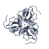 1f7dC 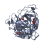 1f7kC 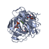 1f7nC 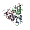 1f7oC 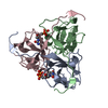 1f7pC 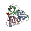 1f7qC C: citing same article ( |
|---|---|
| Similar structure data |


-Links
 PDBj
PDBj


- Assembly
| Deposited unit | 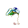
| ||||||||
|---|---|---|---|---|---|---|---|---|---|
| 1 | 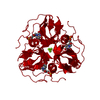
| ||||||||
| Unit cell |
| ||||||||
| Details | Trimer is the functional form of the enzyme |
-Components
| #1: Protein | Mass: 14730.169 Da / Num. of mol.: 1 / Fragment: DUTPASE Source method: isolated from a genetically manipulated source Source: (gene. exp.) Feline immunodeficiency virus / Genus: Lentivirus / Plasmid: FIV-34TF10 / Production host:  Trichoplusia ni (cabbage looper) / References: UniProt: P16088, dUTP diphosphatase Trichoplusia ni (cabbage looper) / References: UniProt: P16088, dUTP diphosphatase |
|---|---|
| #2: Chemical | ChemComp-MG /   Mass: 24.305 Da / Num. of mol.: 1 / Source method: obtained synthetically / Formula: Mg Mass: 24.305 Da / Num. of mol.: 1 / Source method: obtained synthetically / Formula: Mg |
| #3: Chemical | ChemComp-UDP /   Type: RNA linking / Mass: 404.161 Da / Num. of mol.: 1 / Source method: obtained synthetically / Formula: C9H14N2O12P2 / Comment: UDP*YM Type: RNA linking / Mass: 404.161 Da / Num. of mol.: 1 / Source method: obtained synthetically / Formula: C9H14N2O12P2 / Comment: UDP*YM |
| #4: Water | ChemComp-HOH /  Mass: 18.015 Da / Num. of mol.: 54 / Source method: isolated from a natural source / Formula: H2O Mass: 18.015 Da / Num. of mol.: 54 / Source method: isolated from a natural source / Formula: H2O |
-Experimental details
-Experiment
| Experiment | Method: X-RAY DIFFRACTION / Number of used crystals: 1 |
|---|
- Sample preparation
| Crystal | Density Matthews: 2.15 Å3/Da / Density % sol: 42.79 % | ||||||||||||||||||||||||||||||||||||
|---|---|---|---|---|---|---|---|---|---|---|---|---|---|---|---|---|---|---|---|---|---|---|---|---|---|---|---|---|---|---|---|---|---|---|---|---|---|
| Crystal grow | Temperature: 295 K / Method: vapor diffusion, sitting drop / pH: 6.5 Details: 1.25 M sodium citrate. The protein solution was incubated with 25 mM dUDP prior to crystallization experiments., pH 6.5, VAPOR DIFFUSION, SITTING DROP, temperature 295K | ||||||||||||||||||||||||||||||||||||
| Crystal | *PLUS Density % sol: 41 % | ||||||||||||||||||||||||||||||||||||
| Crystal grow | *PLUS pH: 7.5 | ||||||||||||||||||||||||||||||||||||
| Components of the solutions | *PLUS
|
-Data collection
| Diffraction | Mean temperature: 298 K |
|---|---|
| Diffraction source | Source: ROTATING ANODE / Type: SIEMENS / Wavelength: 1.5418 |
| Detector | Type: MARRESEARCH / Detector: IMAGE PLATE / Date: Sep 27, 1998 |
| Radiation | Protocol: SINGLE WAVELENGTH / Monochromatic (M) / Laue (L): M / Scattering type: x-ray |
| Radiation wavelength | Wavelength: 1.5418 Å / Relative weight: 1 |
| Reflection | Resolution: 2.5→50 Å / Num. all: 10960 / Num. obs: 4414 / % possible obs: 96.4 % / Observed criterion σ(F): 0 / Observed criterion σ(I): 0 / Redundancy: 2.5 % / Rmerge(I) obs: 0.104 / Net I/σ(I): 11.1 |
| Reflection shell | Resolution: 2.5→2.59 Å / Rmerge(I) obs: 0.246 / % possible all: 92.5 |
| Reflection | *PLUS Num. measured all: 10960 |
| Reflection shell | *PLUS % possible obs: 92.5 % / Mean I/σ(I) obs: 4.5 |
- Processing
| Refinement | Resolution: 2.5→50 Å / σ(F): 2 /
| ||||||||||||
|---|---|---|---|---|---|---|---|---|---|---|---|---|---|
| Refinement step | Cycle: LAST / Resolution: 2.5→50 Å
| ||||||||||||
| Refine LS restraints |
| ||||||||||||
| Refinement | *PLUS Lowest resolution: 50 Å / σ(F): 2 / % reflection Rfree: 10 % / Rfactor Rfree: 0.266 / Rfactor Rwork: 0.192 | ||||||||||||
| Solvent computation | *PLUS | ||||||||||||
| Displacement parameters | *PLUS | ||||||||||||
| Refine LS restraints | *PLUS
|
