 Movie
Movie Controller


+ Open data

- Basic information
| Entry | Database: PDB / ID: 1a0d | ||||||
|---|---|---|---|---|---|---|---|
| Title | XYLOSE ISOMERASE FROM BACILLUS STEAROTHERMOPHILUS | ||||||
 Components Components | XYLOSE ISOMERASE | ||||||
 Keywords Keywords | KETOLISOMERASE / XYLOSE METABOLISM / GLUCOSE-FRUCTOSE INTERCONVERSION / HYDRIDE TRANSFER / ALPHA-BETA BARREL / METALLOENZYME / THERMOPHILE | ||||||
| Function / homology |  Function and homology information Function and homology informationxylose isomerase / xylose isomerase activity / D-xylose metabolic process / magnesium ion binding / cytoplasm Similarity search - Function | ||||||
| Biological species |   Geobacillus stearothermophilus (bacteria) Geobacillus stearothermophilus (bacteria) | ||||||
| Method |  X-RAY DIFFRACTION / MOLECULAR REPLACEMENT / Resolution: 3 Å X-RAY DIFFRACTION / MOLECULAR REPLACEMENT / Resolution: 3 Å | ||||||
 Authors Authors | Gallay, O. / Chopra, R. / Conti, E. / Brick, P. / Blow, D. | ||||||
 Citation Citation | Journal: To be Published Title: Crystal Structures of Class II Xylose Isomerases from Two Thermophiles and a Hyperthermophile Authors: Gallay, O. / Chopra, R. / Conti, E. / Brick, P. / Jackson, R. / Hartley, B. / Vieille, C. / Zeikus, J.G. / Blow, D. #1: Journal: Biotechnol.Lett. / Year: 1993Title: High Level Expression of a Thermostable Bacillus Xylose (Glucose) Isomerase in Escherichia Coli Authors: Wuxiang, L. / Jeyaseelan, K. | ||||||
| History |
|
- Structure visualization
| Structure viewer | Molecule: MolmilJmol/JSmol |
|---|
- Downloads & links
-Download
| PDBx/mmCIF format | 1a0d.cif.gz | 348.4 KB | Display | PDBx/mmCIF format |
|---|---|---|---|---|
| PDB format | pdb1a0d.ent.gz | 285.4 KB | Display | PDB format |
| PDBx/mmJSON format | 1a0d.json.gz | Tree view | PDBx/mmJSON format | |
| Others |  Other downloads Other downloads |
-Validation report
| Arichive directory | https://data.pdbj.org/pub/pdb/validation_reports/a0/1a0dftp://data.pdbj.org/pub/pdb/validation_reports/a0/1a0d | HTTPS FTP |
|---|
-Related structure data
| Related structure data |  1a0cC  1a0eC  6xiaS S: Starting model for refinement C: citing same article ( |
|---|---|
| Similar structure data |


-Links
 PDBj
PDBj
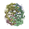
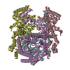
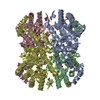
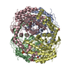
- Assembly
| Deposited unit | 
| ||||||||||||||||
|---|---|---|---|---|---|---|---|---|---|---|---|---|---|---|---|---|---|
| 1 |
| ||||||||||||||||
| Unit cell |
| ||||||||||||||||
| Noncrystallographic symmetry (NCS) | NCS oper:
|
-Components
| #1: Protein | Mass: 50073.395 Da / Num. of mol.: 4 / Source method: isolated from a natural source / Source: (natural) Geobacillus stearothermophilus (bacteria) / Cellular location: CYTOPLASM / Strain: LLD-R / References: UniProt: P54273, xylose isomerase#2: Chemical | ChemComp-MN /   Mass: 54.938 Da / Num. of mol.: 8 / Source method: obtained synthetically / Formula: Mn Mass: 54.938 Da / Num. of mol.: 8 / Source method: obtained synthetically / Formula: Mn#3: Water | ChemComp-HOH / |  Mass: 18.015 Da / Num. of mol.: 240 / Source method: isolated from a natural source / Formula: H2O Mass: 18.015 Da / Num. of mol.: 240 / Source method: isolated from a natural source / Formula: H2OHas protein modification | Y | |
|---|
-Experimental details
-Experiment
| Experiment | Method: X-RAY DIFFRACTION / Number of used crystals: 1 |
|---|
- Sample preparation
| Crystal | Density Matthews: 2.7 Å3/Da / Density % sol: 45 % |
|---|---|
| Crystal grow | Method: vapor diffusion, hanging drop / pH: 6.5 Details: VAPOR DIFFUSION FROM HANGING DROPS (18 DEG C): PROTEIN (A280 22) WAS IN 50 MM TRIS, 10 MM MNCL2, PH 7.5; RESERVOIR SOLUTION WAS 10% PEG, 100 MM LICL, 100 MM MES, PH 6.3; DROPS WERE FORMED ...Details: VAPOR DIFFUSION FROM HANGING DROPS (18 DEG C): PROTEIN (A280 22) WAS IN 50 MM TRIS, 10 MM MNCL2, PH 7.5; RESERVOIR SOLUTION WAS 10% PEG, 100 MM LICL, 100 MM MES, PH 6.3; DROPS WERE FORMED FROM EQUAL PARTS OF PROTEIN AND RESERVOIR SOLUTIONS., pH 6.5, vapor diffusion - hanging drop PH range: 6.3 - 7.5 |
-Data collection
| Diffraction | Mean temperature: 293 K |
|---|---|
| Diffraction source | Source: ROTATING ANODE / Type: ELLIOTT GX-21 / Wavelength: 1.5418 |
| Detector | Type: ENRAF-NONIUS FAST / Detector: DIFFRACTOMETER / Date: Feb 1, 1993 / Details: DUAL SLITS, COLLIMATOR |
| Radiation | Monochromator: GRAPHITE(002) / Monochromatic (M) / Laue (L): M / Scattering type: x-ray |
| Radiation wavelength | Wavelength: 1.5418 Å / Relative weight: 1 |
| Reflection | Resolution: 3→33 Å / Num. obs: 35985 / % possible obs: 89.6 % / Redundancy: 1.7 % / Biso Wilson estimate: 49.8 Å2 / Rmerge(I) obs: 0.1 / Net I/σ(I): 7.1 |
| Reflection shell | Resolution: 3→3.16 Å / Redundancy: 1.5 % / Rmerge(I) obs: 0.23 / Mean I/σ(I) obs: 3.1 / % possible all: 62.2 |
- Processing
| Software |
| ||||||||||||||||||||||||||||||||||||||||||||||||||||||||||||
|---|---|---|---|---|---|---|---|---|---|---|---|---|---|---|---|---|---|---|---|---|---|---|---|---|---|---|---|---|---|---|---|---|---|---|---|---|---|---|---|---|---|---|---|---|---|---|---|---|---|---|---|---|---|---|---|---|---|---|---|---|---|
| Refinement | Method to determine structure: MOLECULAR REPLACEMENT Starting model: PDB ENTRY 6XIA Resolution: 3→20 Å / Rfactor Rfree error: 0.005 / Cross valid method: A POSTERIORI Details: OCCUPANCIES OF THE MN CATIONS AND WATER HOH 501 WERE REFINED, SIMULTANEOUSLY WITH TEMPERATURE FACTORS FOR ALL ATOMS, USING THE GROUP B COMMAND IN X-PLOR. THESE OCCUPANCIES WERE THEN FIXED AT ...Details: OCCUPANCIES OF THE MN CATIONS AND WATER HOH 501 WERE REFINED, SIMULTANEOUSLY WITH TEMPERATURE FACTORS FOR ALL ATOMS, USING THE GROUP B COMMAND IN X-PLOR. THESE OCCUPANCIES WERE THEN FIXED AT ROUNDED VALUES FOR THE SUBSEQUENT REFINEMENT PROTOCOL. DISORDERED SIDE CHAINS WERE NOT INCLUDED IN REFINEMENT.
| ||||||||||||||||||||||||||||||||||||||||||||||||||||||||||||
| Displacement parameters | Biso mean: 19.1 Å2 | ||||||||||||||||||||||||||||||||||||||||||||||||||||||||||||
| Refinement step | Cycle: LAST / Resolution: 3→20 Å
| ||||||||||||||||||||||||||||||||||||||||||||||||||||||||||||
| Refine LS restraints |
| ||||||||||||||||||||||||||||||||||||||||||||||||||||||||||||
| LS refinement shell | Resolution: 3→3.14 Å / Rfactor Rfree error: 0.025 / Total num. of bins used: 8
| ||||||||||||||||||||||||||||||||||||||||||||||||||||||||||||
| Xplor file |
|
