Movie
 Controller


[English] 日本語
 Yorodumi
- PDB-12tr: 2 molecules of Fab NORO-320 bound to C-D VP1 dimer of GII.4 Syd V... -
+ Open data

- Basic information
| Entry | Database: PDB / ID: 12tr | |||||||||
|---|---|---|---|---|---|---|---|---|---|---|
| Title | 2 molecules of Fab NORO-320 bound to C-D VP1 dimer of GII.4 Syd VLP (T=4) | |||||||||
 Components |
| |||||||||
 Keywords | VIRUS LIKE PARTICLE / Norovirus / Sydney / VLP / Capsid / Antibody | |||||||||
| Function / homology | Calicivirus coat protein C-terminal / Calicivirus coat protein C-terminal / Calicivirus coat protein / Calicivirus coat protein / Picornavirus/Calicivirus coat protein / Viral coat protein subunit / host cell cytoplasm / VP1 Function and homology information | |||||||||
| Biological species |  Norovirus Hu/GII.4/Sydney/NSW0514/2012/AU  Homo sapiens (human) | |||||||||
| Method | ELECTRON MICROSCOPY / single particle reconstruction / cryo EM / Resolution: 6.28 Å | |||||||||
 Authors | Apostol, C.V. / Anish, R. / Pham, S. / Prasad, B.V.V. | |||||||||
| Funding support |  United States, 1items
| |||||||||
 Citation | Journal: Res Sq / Year: 2026 Title: Symmetry-breaking antibody binding reveals extensive capsid malleability in pandemic human norovirus. Authors: Carmen V Apostol / Ramakrishnan Anish / Son Pham / Khalil Ettayebi / Janam Dave / Frederick H Neill / Marina Bok / Banumathi Sankaran / Sue Crawford / Robert L Atmar / James E Crowe / ...Authors: Carmen V Apostol / Ramakrishnan Anish / Son Pham / Khalil Ettayebi / Janam Dave / Frederick H Neill / Marina Bok / Banumathi Sankaran / Sue Crawford / Robert L Atmar / James E Crowe / Viviana Parreño / Mary K Estes / B V Venkataram Prasad /  Abstract: Virus capsids are metastable assemblies with an exquisite balance between stability and flexibility, obligatory for entry, infectivity, and immune escape. Yet, the extent to which human viruses ...Virus capsids are metastable assemblies with an exquisite balance between stability and flexibility, obligatory for entry, infectivity, and immune escape. Yet, the extent to which human viruses exploit capsid plasticity and how that consequently shapes antigenic landscapes remains poorly understood. Human noroviruses (HuNoVs) are the leading cause of acute viral gastroenteritis worldwide. GII.4 HuNoVs have been evolving epochally, driving most global pandemics, and since 2012, GII.4 Sydney has been the dominant variant. The major capsid protein VP1 is central to infectivity and immune recognition, and VP1-based virus-like particles (VLPs) are considered promising vaccine candidates. Yet, how the icosahedral HuNoV capsid responds to antibody pressure at the atomic level is still unclear. Here, we present the first atomic structure of GII.4 Sydney capsid determined by cryo-EM, revealing the structural features that likely contribute to its sustained dominance. Using focused refinement, we uncovered the remarkable capsid plasticity in response to symmetry-breaking binding of neutralizing and non-neutralizing antibodies. VP1 adopts 11 distinct conformational modalities to accommodate asymmetric antibody binding, and antibody-disrupted VLPs reassemble into smaller particles with five additional VP1 states. This previously unrecognized extent of conformational adaptability provides key insights for designing next-generation VLP- or mRNA-based vaccines targeting HuNoV infections. | |||||||||
| History |
|
- Structure visualization
| Structure viewer | Molecule: MolmilJmol/JSmol |
|---|
- Downloads & links
-Download
| PDBx/mmCIF format | 12tr.cif.gz | 321 KB | Display | PDBx/mmCIF format |
|---|---|---|---|---|
| PDB format | pdb12tr.ent.gz | 260.6 KB | Display | PDB format |
| PDBx/mmJSON format | 12tr.json.gz | Tree view | PDBx/mmJSON format | |
| Others |  Other downloads |
-Validation report
| Arichive directory | https://data.pdbj.org/pub/pdb/validation_reports/2t/12trftp://data.pdbj.org/pub/pdb/validation_reports/2t/12tr | HTTPS FTP |
|---|
-Related structure data
| Related structure data |  76751MC  12pgC  12qzC  12tfC  12tpC  12tqC  12tsC  12ttC  12tuC  12tvC  12twC  12txC  12tyC  12tzC  12uaC  12ubC  12ucC  12udC  12ueC  13kfC  13kgC M: map data used to model this data C: citing same article ( |
|---|---|
| Similar structure data |


-Links
 PDBj
- Assembly
| Deposited unit | 
|
|---|---|
| 1 |
|
-Components
| #1: Protein | Mass: 59157.246 Da / Num. of mol.: 2 Source method: isolated from a genetically manipulated source Source: (gene. exp.) Norovirus Hu/GII.4/Sydney/NSW0514/2012/AUProduction host:   Spodoptera frugiperda (fall armyworm) / References: UniProt: K4LM89 #2: Antibody | Mass: 25202.338 Da / Num. of mol.: 2 Source method: isolated from a genetically manipulated source Source: (gene. exp.) Homo sapiens (human) / Production host:   Cricetulus griseus (Chinese hamster) #3: Antibody | Mass: 23905.609 Da / Num. of mol.: 2 Source method: isolated from a genetically manipulated source Source: (gene. exp.) Homo sapiens (human) / Production host: Cricetulus griseus (Chinese hamster)Has protein modification | Y | |
|---|
-Experimental details
-Experiment
| Experiment | Method: ELECTRON MICROSCOPY |
|---|---|
| EM experiment | Aggregation state: PARTICLE / 3D reconstruction method: single particle reconstruction |
- Sample preparation
| Component | Name: Norovirus / Type: VIRUS / Entity ID: all / Source: RECOMBINANT |
|---|---|
| Source (natural) | Organism: Norovirus |
| Source (recombinant) | Organism: Spodoptera frugiperda (fall armyworm) |
| Details of virus | Empty: YES / Enveloped: YES / Isolate: STRAIN / Type: VIRUS-LIKE PARTICLE |
| Natural host | Organism: Norovirus Hu/GII.4/Sydney/NSW0514/2012/AU |
| Virus shell | Name: VP1 / Diameter: 420 nm / Triangulation number (T number): 3 |
| Buffer solution | pH: 6 / Details: 1X PBS |
| Specimen | Embedding applied: NO / Shadowing applied: NO / Staining applied: NO / Vitrification applied: YES |
| Specimen support | Grid material: COPPER / Grid mesh size: 200 divisions/in. / Grid type: Quantifoil R1.2/1.3 |
| Vitrification | Instrument: FEI VITROBOT MARK IV / Cryogen name: ETHANE / Humidity: 100 % / Chamber temperature: 291 K |
- Electron microscopy imaging
| Experimental equipment | 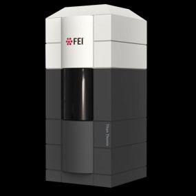 Model: Titan Krios / Image courtesy: FEI Company |
|---|---|
| Microscopy | Model: TFS KRIOS |
| Electron gun | Electron source:  FIELD EMISSION GUN / Accelerating voltage: 300 kV / Illumination mode: FLOOD BEAM |
| Electron lens | Mode: BRIGHT FIELD / Nominal magnification: 130000 X / Nominal defocus max: 2500 nm / Nominal defocus min: 500 nm / Cs: 2.7 mm / C2 aperture diameter: 70 µm |
| Specimen holder | Cryogen: NITROGEN |
| Image recording | Electron dose: 50 e/Å2 / Film or detector model: GATAN K2 SUMMIT (4k x 4k) / Num. of grids imaged: 1 / Num. of real images: 3290 Details: Images were collected in movie mode at 35 frames per second |
| EM imaging optics | Energyfilter name: GIF Bioquantum |
- Processing
| EM software |
| ||||||||||||||||||||||||||||||||
|---|---|---|---|---|---|---|---|---|---|---|---|---|---|---|---|---|---|---|---|---|---|---|---|---|---|---|---|---|---|---|---|---|---|
| CTF correction | Type: NONE | ||||||||||||||||||||||||||||||||
| 3D reconstruction | Resolution: 6.28 Å / Resolution method: FSC 0.143 CUT-OFF / Num. of particles: 22827 / Symmetry type: POINT | ||||||||||||||||||||||||||||||||
| Refinement | Highest resolution: 6.28 Å Stereochemistry target values: REAL-SPACE (WEIGHTED MAP SUM AT ATOM CENTERS) | ||||||||||||||||||||||||||||||||
| Refine LS restraints |
|