Movie
 Controller


+ Open data

- Basic information
| Entry | Database: PDB / ID: 12pg | ||||||
|---|---|---|---|---|---|---|---|
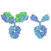
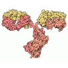
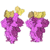
| Title | GII.3 P-domain in complex with M6 Nanobody | ||||||
 Components |
| ||||||
 Keywords | VIRAL PROTEIN / P-domain / nanobody / Norovirus | ||||||
| Function / homology | :  Function and homology information | ||||||
| Biological species |  Norovirus Hu/Texas/TCH04-577/2004/US   Lama glama (llama) | ||||||
| Method |  X-RAY DIFFRACTION / SYNCHROTRON / MOLECULAR REPLACEMENT / Resolution: 2.2 Å | ||||||
 Authors | Apostol, C.V. / Anish, R. / Pham, S. / Sankaran, B. / Prasad, B.V.V. | ||||||
| Funding support |  United States, 1items
| ||||||
 Citation | Journal: Res Sq / Year: 2026 Title: Symmetry-breaking antibody binding reveals extensive capsid malleability in pandemic human norovirus. Authors: Carmen V Apostol / Ramakrishnan Anish / Son Pham / Khalil Ettayebi / Janam Dave / Frederick H Neill / Marina Bok / Banumathi Sankaran / Sue Crawford / Robert L Atmar / James E Crowe / ...Authors: Carmen V Apostol / Ramakrishnan Anish / Son Pham / Khalil Ettayebi / Janam Dave / Frederick H Neill / Marina Bok / Banumathi Sankaran / Sue Crawford / Robert L Atmar / James E Crowe / Viviana Parreño / Mary K Estes / B V Venkataram Prasad /  Abstract: Virus capsids are metastable assemblies with an exquisite balance between stability and flexibility, obligatory for entry, infectivity, and immune escape. Yet, the extent to which human viruses ...Virus capsids are metastable assemblies with an exquisite balance between stability and flexibility, obligatory for entry, infectivity, and immune escape. Yet, the extent to which human viruses exploit capsid plasticity and how that consequently shapes antigenic landscapes remains poorly understood. Human noroviruses (HuNoVs) are the leading cause of acute viral gastroenteritis worldwide. GII.4 HuNoVs have been evolving epochally, driving most global pandemics, and since 2012, GII.4 Sydney has been the dominant variant. The major capsid protein VP1 is central to infectivity and immune recognition, and VP1-based virus-like particles (VLPs) are considered promising vaccine candidates. Yet, how the icosahedral HuNoV capsid responds to antibody pressure at the atomic level is still unclear. Here, we present the first atomic structure of GII.4 Sydney capsid determined by cryo-EM, revealing the structural features that likely contribute to its sustained dominance. Using focused refinement, we uncovered the remarkable capsid plasticity in response to symmetry-breaking binding of neutralizing and non-neutralizing antibodies. VP1 adopts 11 distinct conformational modalities to accommodate asymmetric antibody binding, and antibody-disrupted VLPs reassemble into smaller particles with five additional VP1 states. This previously unrecognized extent of conformational adaptability provides key insights for designing next-generation VLP- or mRNA-based vaccines targeting HuNoV infections. | ||||||
| History |
|
- Structure visualization
| Structure viewer | Molecule: MolmilJmol/JSmol |
|---|
- Downloads & links
-Download
| PDBx/mmCIF format | 12pg.cif.gz | 172.2 KB | Display | PDBx/mmCIF format |
|---|---|---|---|---|
| PDB format | pdb12pg.ent.gz | 133.6 KB | Display | PDB format |
| PDBx/mmJSON format | 12pg.json.gz | Tree view | PDBx/mmJSON format | |
| Others |  Other downloads |
-Validation report
| Arichive directory | https://data.pdbj.org/pub/pdb/validation_reports/2p/12pgftp://data.pdbj.org/pub/pdb/validation_reports/2p/12pg | HTTPS FTP |
|---|
-Related structure data
| Related structure data |  12qzC  12tfC  12tpC  12tqC  12trC  12tsC  12ttC  12tuC  12tvC  12twC  12txC  12tyC  12tzC  12uaC  12ubC  12ucC  12udC  12ueC  13kfC  13kgC C: citing same article ( |
|---|---|
| Similar structure data |


-Links
 PDBj
- Assembly
| Deposited unit | 
| ||||||||||||
|---|---|---|---|---|---|---|---|---|---|---|---|---|---|
| 1 | 
| ||||||||||||
| Unit cell |
| ||||||||||||
| Components on special symmetry positions |
|
-Components
| #1: Protein | Mass: 35617.637 Da / Num. of mol.: 1 Source method: isolated from a genetically manipulated source Source: (gene. exp.) Norovirus Hu/Texas/TCH04-577/2004/USProduction host:   Escherichia coli 'BL21-Gold(DE3)pLysS AG' (bacteria) References: UniProt: B2DD27 |
|---|---|
| #2: Antibody | Mass: 13308.611 Da / Num. of mol.: 1 Source method: isolated from a genetically manipulated source Source: (gene. exp.) Lama glama (llama)Production host: Escherichia coli 'BL21-Gold(DE3)pLysS AG' (bacteria) |
| #3: Chemical | ChemComp-CL /   Mass: 35.453 Da / Num. of mol.: 1 / Source method: obtained synthetically / Formula: Cl / Feature type: SUBJECT OF INVESTIGATION |
| #4: Water | ChemComp-HOH /  Mass: 18.015 Da / Num. of mol.: 182 / Source method: isolated from a natural source / Formula: H2O |
| Has ligand of interest | Y |
| Has protein modification | Y |
-Experimental details
-Experiment
| Experiment | Method: X-RAY DIFFRACTION / Number of used crystals: 1 |
|---|
- Sample preparation
| Crystal | Density Matthews: 2.54 Å3/Da / Density % sol: 51.55 % |
|---|---|
| Crystal grow | Temperature: 298 K / Method: vapor diffusion, hanging drop Details: 300mM succinic acid 100mM citric acid NaOH, pH 3.5 22.5% PurePEGs cocktail 10mM phenol |
-Data collection
| Diffraction | Mean temperature: 100 K / Serial crystal experiment: N |
|---|---|
| Diffraction source | Source: SYNCHROTRON / Site: ALS / Beamline: 8.2.2 / Wavelength: 1.00002 Å |
| Detector | Type: DECTRIS PILATUS3 S 2M / Detector: PIXEL / Date: Dec 21, 2023 |
| Radiation | Protocol: SINGLE WAVELENGTH / Monochromatic (M) / Laue (L): M / Scattering type: x-ray |
| Radiation wavelength | Wavelength: 1.00002 Å / Relative weight: 1 |
| Reflection | Resolution: 2.2→45.24 Å / Num. obs: 26770 / % possible obs: 100 % / Redundancy: 30 % / Biso Wilson estimate: 33.23 Å2 / Rmerge(I) obs: 0.139 / Net I/σ(I): 14.8 |
| Reflection shell | Resolution: 2.2→2.24 Å / Rmerge(I) obs: 0.785 / Num. unique obs: 1307 |
- Processing
| Software |
| |||||||||||||||||||||||||||||||||||||||||||||||||||||||||||||||||||||||||||||
|---|---|---|---|---|---|---|---|---|---|---|---|---|---|---|---|---|---|---|---|---|---|---|---|---|---|---|---|---|---|---|---|---|---|---|---|---|---|---|---|---|---|---|---|---|---|---|---|---|---|---|---|---|---|---|---|---|---|---|---|---|---|---|---|---|---|---|---|---|---|---|---|---|---|---|---|---|---|---|
| Refinement | Method to determine structure: MOLECULAR REPLACEMENT / Resolution: 2.2→45.24 Å / SU ML: 0.2222 / Cross valid method: FREE R-VALUE / σ(F): 1.34 / Phase error: 21.9052 Stereochemistry target values: GeoStd + Monomer Library + CDL v1.2
| |||||||||||||||||||||||||||||||||||||||||||||||||||||||||||||||||||||||||||||
| Solvent computation | Shrinkage radii: 0.9 Å / VDW probe radii: 1.1 Å / Solvent model: FLAT BULK SOLVENT MODEL | |||||||||||||||||||||||||||||||||||||||||||||||||||||||||||||||||||||||||||||
| Displacement parameters | Biso mean: 41.45 Å2 | |||||||||||||||||||||||||||||||||||||||||||||||||||||||||||||||||||||||||||||
| Refinement step | Cycle: LAST / Resolution: 2.2→45.24 Å
| |||||||||||||||||||||||||||||||||||||||||||||||||||||||||||||||||||||||||||||
| Refine LS restraints |
| |||||||||||||||||||||||||||||||||||||||||||||||||||||||||||||||||||||||||||||
| LS refinement shell |
|