 Movie
Movie Controller


+ Open data

- Basic information
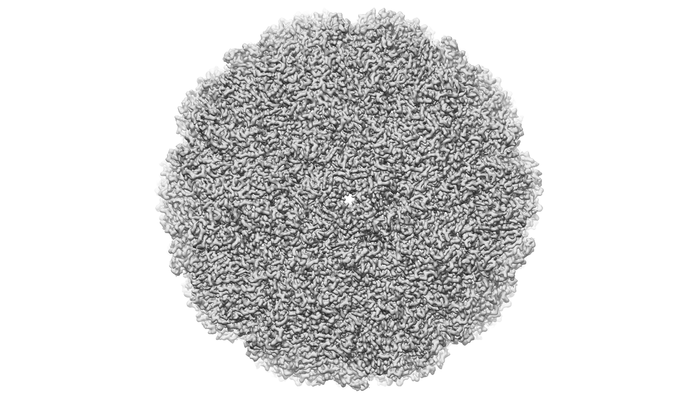
| Entry | Database: EMDB / ID: EMD-9674 | |||||||||
|---|---|---|---|---|---|---|---|---|---|---|
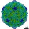
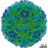
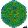
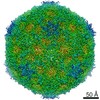
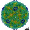
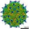
| Title | Cryo-EM structure of CV-A10 mature virion | |||||||||
 Map data Map data | ||||||||||
 Sample Sample |
| |||||||||
 Keywords Keywords | Coxsackievirus A10 / Mature virion / VIRAL PROTEIN | |||||||||
| Function / homology |  Function and homology information Function and homology informationsymbiont genome entry into host cell via pore formation in plasma membrane / viral capsid / host cell cytoplasm / symbiont-mediated suppression of host gene expression / virion attachment to host cell / structural molecule activity Similarity search - Function | |||||||||
| Biological species |  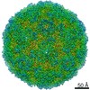 Coxsackievirus A10 Coxsackievirus A10 | |||||||||
| Method | single particle reconstruction / cryo EM / Resolution: 2.84 Å | |||||||||
 Authors Authors | Chen JH / Ye XH | |||||||||
 Citation Citation | Journal: Cell Discov / Year: 2019 Title: Coxsackievirus A10 atomic structure facilitating the discovery of a broad-spectrum inhibitor against human enteroviruses. Authors: Jinhuan Chen / Xiaohua Ye / Xue-Yang Zhang / Zhengdan Zhu / Xiang Zhang / Zhijian Xu / Zhanyu Ding / Gang Zou / Qingwei Liu / Liangliang Kong / Wen Jiang / Weiliang Zhu / Yao Cong / Zhong Huang /   Abstract: Coxsackievirus A10 (CV-A10) belongs to the species A and is a causative agent of hand, foot, and mouth disease. Here we present cryo-EM structures of CV-A10 mature virion and native empty particle ...Coxsackievirus A10 (CV-A10) belongs to the species A and is a causative agent of hand, foot, and mouth disease. Here we present cryo-EM structures of CV-A10 mature virion and native empty particle (NEP) at 2.84 and 3.12 Å, respectively. Our CV-A10 mature virion structure reveals a density corresponding to a lipidic pocket factor of 18 carbon atoms in the hydrophobic pocket formed within viral protein 1. By structure-guided high-throughput drug screening and subsequent verification in cell-based infection-inhibition assays, we identified four compounds that inhibited CV-A10 infection in vitro. These compounds represent a new class of anti-enteroviral drug leads. Notably, one of the compounds, ICA135, also exerted broad-spectrum inhibitory effects on a number of representative viruses from all four species (A-D) of human enteroviruses. Our findings should facilitate the development of broadly effective drugs and vaccines for enterovirus infections. | |||||||||
| History |
|
- Structure visualization
| Movie |
Movie viewer |
|---|---|
| Structure viewer | EM map: SurfViewMolmilJmol/JSmol |
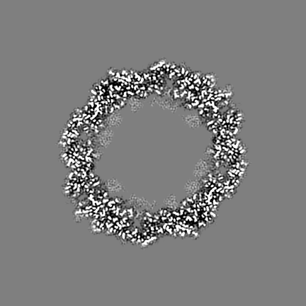
| Supplemental images |
- Downloads & links
-EMDB archive
| Map data | emd_9674.map.gz | 41.3 MB | EMDB map data format | |
|---|---|---|---|---|
| Header (meta data) | emd-9674-v30.xmlemd-9674.xml | 15.3 KB 15.3 KB | Display Display | EMDB header |
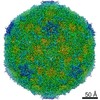
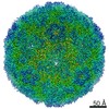
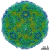
| Images |  emd_9674.png emd_9674.png | 109.9 KB | ||
| Filedesc metadata | emd-9674.cif.gz | 6.3 KB | ||
| Archive directory |  http://ftp.pdbj.org/pub/emdb/structures/EMD-9674ftp://ftp.pdbj.org/pub/emdb/structures/EMD-9674 http://ftp.pdbj.org/pub/emdb/structures/EMD-9674ftp://ftp.pdbj.org/pub/emdb/structures/EMD-9674 | HTTPS FTP |
-Related structure data
| Related structure data | 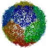 6iijMC 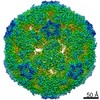 9675C 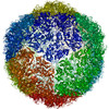 6iioC M: atomic model generated by this map C: citing same article ( |
|---|---|
| Similar structure data |

-Links
| EMDB pages | EMDB (EBI/PDBe) / EMDataResource |
|---|---|
| Related items in Molecule of the Month |

-Map
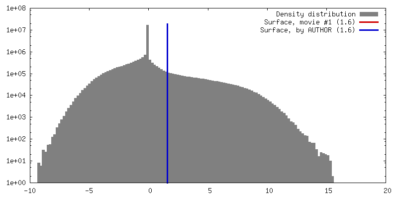
| File | Download / File: emd_9674.map.gz / Format: CCP4 / Size: 307.5 MB / Type: IMAGE STORED AS FLOATING POINT NUMBER (4 BYTES) | ||||||||||||||||||||||||||||||||||||||||||||||||||||||||||||||||||||
|---|---|---|---|---|---|---|---|---|---|---|---|---|---|---|---|---|---|---|---|---|---|---|---|---|---|---|---|---|---|---|---|---|---|---|---|---|---|---|---|---|---|---|---|---|---|---|---|---|---|---|---|---|---|---|---|---|---|---|---|---|---|---|---|---|---|---|---|---|---|
| Projections & slices | Image control
Images are generated by Spider. | ||||||||||||||||||||||||||||||||||||||||||||||||||||||||||||||||||||
| Voxel size | X=Y=Z: 1.005 Å | ||||||||||||||||||||||||||||||||||||||||||||||||||||||||||||||||||||
| Density |
| ||||||||||||||||||||||||||||||||||||||||||||||||||||||||||||||||||||
| Symmetry | Space group: 1 | ||||||||||||||||||||||||||||||||||||||||||||||||||||||||||||||||||||
| Details | EMDB XML:
CCP4 map header:
| ||||||||||||||||||||||||||||||||||||||||||||||||||||||||||||||||||||
 Z (Sec.)
 Y (Row.)
 X (Col.)


-Supplemental data
- Sample components
-Entire : Coxsackievirus A10
| Entire | Name: Coxsackievirus A10 |
|---|---|
| Components |
|
-Supramolecule #1: Coxsackievirus A10
| Supramolecule | Name: Coxsackievirus A10 / type: virus / ID: 1 / Parent: 0 / Macromolecule list: #1-#4 / NCBI-ID: 42769 / Sci species name: Coxsackievirus A10 / Virus type: VIRION / Virus isolate: STRAIN / Virus enveloped: No / Virus empty: No |
|---|
-Macromolecule #1: VP1
| Macromolecule | Name: VP1 / type: protein_or_peptide / ID: 1 / Number of copies: 1 / Enantiomer: LEVO |
|---|---|
| Source (natural) | Organism: Coxsackievirus A10 |
| Molecular weight | Theoretical: 33.088219 KDa |
| Sequence | String: GDPVEDIIHD ALGNTARRAI SGATNVESAA DTTPSSHRLE TGRVPALQAA ETGATSNATD ENMIETRCVI NRNGVLETTI NHFFSRSGL VGVVNLTDGG DTTGYATWDI DIMGFVQLRR KCEMFTYMRF NAEFTFVTTT KSGEARPYML QYMYVPPGAP K PTGRDAFQ ...String: GDPVEDIIHD ALGNTARRAI SGATNVESAA DTTPSSHRLE TGRVPALQAA ETGATSNATD ENMIETRCVI NRNGVLETTI NHFFSRSGL VGVVNLTDGG DTTGYATWDI DIMGFVQLRR KCEMFTYMRF NAEFTFVTTT KSGEARPYML QYMYVPPGAP K PTGRDAFQ WQTATNPSVF VKLTDPPAQV SVPFMSPASA YQWFYDGYPT FGQHPETSNT TYGLCPNNMM GTFAVRVVSR EA SQLKLQT RVYMKLKHVR AWVPRPIRSQ PYLLKNFPNY DSSKITNSAR DRSSIKQANM UniProtKB: Genome polyprotein |
-Macromolecule #2: VP2
| Macromolecule | Name: VP2 / type: protein_or_peptide / ID: 2 / Number of copies: 1 / Enantiomer: LEVO |
|---|---|
| Source (natural) | Organism: Coxsackievirus A10 |
| Molecular weight | Theoretical: 27.741023 KDa |
| Sequence | String: SPSVEACGYS DRVAQLTVGN SSITTQEAAN IVLAYGEWPE YCPDTDATAV DKPTRPDVSV NRFYTLDSKM WQENSTGWYW KFPDVLNKT GVFGQNAQFH YLYRSGFCLH VQCNASKFHQ GALLVAVIPE FVIAGRGSNT KPNEAPHPGF TTTFPGTTGA T FHDPYVLD ...String: SPSVEACGYS DRVAQLTVGN SSITTQEAAN IVLAYGEWPE YCPDTDATAV DKPTRPDVSV NRFYTLDSKM WQENSTGWYW KFPDVLNKT GVFGQNAQFH YLYRSGFCLH VQCNASKFHQ GALLVAVIPE FVIAGRGSNT KPNEAPHPGF TTTFPGTTGA T FHDPYVLD SGVPLSQALI YPHQWVNLRT NNCATVIVPY INAVPFDSAI NHSNFGLVVV PVSPLKYSSG ATTAIPITIT IA PLNSEFG GLRQAVSQ UniProtKB: Genome polyprotein |
-Macromolecule #3: VP3
| Macromolecule | Name: VP3 / type: protein_or_peptide / ID: 3 / Number of copies: 1 / Enantiomer: LEVO |
|---|---|
| Source (natural) | Organism: Coxsackievirus A10 |
| Molecular weight | Theoretical: 26.187623 KDa |
| Sequence | String: GIPAELRPGT NQFLTTDDDT AAPILPGFTP TPTIHIPGEV HSLLELCRVE TILEVNNTTE ATGLTRLLIP VSSQNKADEL CAAFMVDPG RIGPWQSTLV GQICRYYTQW SGSLKVTFMF TGSFMATGKM LVAYSPPGSA QPANRETAML GTHVIWDFGL Q SSVSLVIP ...String: GIPAELRPGT NQFLTTDDDT AAPILPGFTP TPTIHIPGEV HSLLELCRVE TILEVNNTTE ATGLTRLLIP VSSQNKADEL CAAFMVDPG RIGPWQSTLV GQICRYYTQW SGSLKVTFMF TGSFMATGKM LVAYSPPGSA QPANRETAML GTHVIWDFGL Q SSVSLVIP WISNTHFRTA KTGGNYDYYT AGVVTLWYQT NYVVPPETPG EAYIIAMGAA QDNFTLKICK DTDEVTQQAV LQ UniProtKB: Genome polyprotein |
-Macromolecule #4: VP4
| Macromolecule | Name: VP4 / type: protein_or_peptide / ID: 4 / Number of copies: 1 / Enantiomer: LEVO |
|---|---|
| Source (natural) | Organism: Coxsackievirus A10 |
| Molecular weight | Theoretical: 7.464104 KDa |
| Sequence | String: MGAQVSTQKS GSHETGNVAT GGSTINFTNI NYYKDSYAAS ATRQDFTQDP KKFTQPVLDS IRELSAPLN UniProtKB: Genome polyprotein |
-Macromolecule #5: SPHINGOSINE
| Macromolecule | Name: SPHINGOSINE / type: ligand / ID: 5 / Number of copies: 1 / Formula: SPH |
|---|---|
| Molecular weight | Theoretical: 299.492 Da |
| Chemical component information |  ChemComp-SPH: |
-Experimental details
-Structure determination
| Method | cryo EM |
|---|---|
 Processing Processing | single particle reconstruction |
| Aggregation state | particle |
-Sample preparation
| Buffer | pH: 7.4 |
|---|---|
| Vitrification | Cryogen name: NITROGEN |
- Electron microscopy
| Microscope | FEI TITAN KRIOS |
|---|---|
| Image recording | Film or detector model: GATAN K2 SUMMIT (4k x 4k) / Average electron dose: 8.0 e/Å2 |
| Electron beam | Acceleration voltage: 300 kV / Electron source:  FIELD EMISSION GUN FIELD EMISSION GUN |
| Electron optics | Illumination mode: OTHER / Imaging mode: BRIGHT FIELD |
| Experimental equipment |  Model: Titan Krios / Image courtesy: FEI Company |
