 Movie
Movie Controller


+ Open data

- Basic information
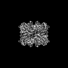
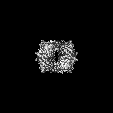
| Entry | Database: EMDB / ID: EMD-9380 | |||||||||
|---|---|---|---|---|---|---|---|---|---|---|
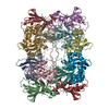
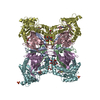
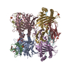
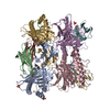
| Title | cryoEM structure of the truncated HIV-1 Vif/CBFbeta/A3F complex | |||||||||
 Map data Map data | ||||||||||
 Sample Sample |
| |||||||||
 Keywords Keywords | Human antiviral restriction factor / HIV viral protein / ANTIVIRAL PROTEIN | |||||||||
| Function / homology |  Function and homology information Function and homology informationRUNX3 regulates RUNX1-mediated transcription / apolipoprotein B mRNA editing enzyme complex / RUNX1 regulates transcription of genes involved in BCR signaling / RUNX1 regulates transcription of genes involved in interleukin signaling / RUNX2 regulates bone development / core-binding factor complex / RUNX1 regulates expression of components of tight junctions / positive regulation of CD8-positive, alpha-beta T cell differentiation / RUNX2 regulates chondrocyte maturation / base conversion or substitution editing ...RUNX3 regulates RUNX1-mediated transcription / apolipoprotein B mRNA editing enzyme complex / RUNX1 regulates transcription of genes involved in BCR signaling / RUNX1 regulates transcription of genes involved in interleukin signaling / RUNX2 regulates bone development / core-binding factor complex / RUNX1 regulates expression of components of tight junctions / positive regulation of CD8-positive, alpha-beta T cell differentiation / RUNX2 regulates chondrocyte maturation / base conversion or substitution editing / single-stranded DNA cytosine deaminase / negative regulation of CD4-positive, alpha-beta T cell differentiation / negative regulation of single stranded viral RNA replication via double stranded DNA intermediate / DNA cytosine deamination / cytidine to uridine editing / clearance of foreign intracellular DNA / negative regulation of viral process / cytidine deaminase activity / RUNX1 and FOXP3 control the development of regulatory T lymphocytes (Tregs) / lymphocyte differentiation / RUNX2 regulates genes involved in cell migration / Transcriptional regulation by RUNX2 / RUNX2 regulates genes involved in differentiation of myeloid cells / RUNX1 regulates transcription of genes involved in differentiation of keratinocytes / positive regulation of gene expression via chromosomal CpG island demethylation / transposable element silencing / RUNX3 Regulates Immune Response and Cell Migration / myeloid cell differentiation / definitive hemopoiesis / RUNX1 regulates transcription of genes involved in differentiation of myeloid cells / Regulation of RUNX1 Expression and Activity / RUNX1 regulates transcription of genes involved in WNT signaling / RUNX1 regulates estrogen receptor mediated transcription / negative regulation of viral genome replication / RUNX2 regulates osteoblast differentiation / RUNX1 interacts with co-factors whose precise effect on RUNX1 targets is not known / RUNX3 regulates p14-ARF / viral life cycle / positive regulation of defense response to virus by host / cell maturation / antiviral innate immune response / P-body / RUNX1 regulates genes involved in megakaryocyte differentiation and platelet function / Regulation of RUNX3 expression and activity / virion component / Transcriptional regulation of granulopoiesis / protein polyubiquitination / osteoblast differentiation / Regulation of RUNX2 expression and activity / RUNX1 regulates transcription of genes involved in differentiation of HSCs / transcription by RNA polymerase II / Estrogen-dependent gene expression / sequence-specific DNA binding / defense response to virus / host cell cytoplasm / transcription coactivator activity / ribonucleoprotein complex / innate immune response / regulation of transcription by RNA polymerase II / host cell plasma membrane / negative regulation of transcription by RNA polymerase II / positive regulation of transcription by RNA polymerase II / RNA binding / zinc ion binding / nucleoplasm / membrane / identical protein binding / nucleus / cytoplasm Similarity search - Function | |||||||||
| Biological species |   Human immunodeficiency virus 1 / Human immunodeficiency virus 1 /  Homo sapiens (human) Homo sapiens (human) | |||||||||
| Method | single particle reconstruction / cryo EM / Resolution: 3.9 Å | |||||||||
 Authors Authors | Hu Y / Xiong Y | |||||||||
| Funding support |  United States, 1 items United States, 1 items
| |||||||||
 Citation Citation | Journal: Nat Struct Mol Biol / Year: 2019 Title: Structural basis of antagonism of human APOBEC3F by HIV-1 Vif. Authors: Yingxia Hu / Belete A Desimmie / Henry C Nguyen / Samantha J Ziegler / Tat Cheung Cheng / John Chen / Jia Wang / Hongwei Wang / Kai Zhang / Vinay K Pathak / Yong Xiong /   Abstract: HIV-1 virion infectivity factor (Vif) promotes degradation of the antiviral APOBEC3 (A3) proteins through the host ubiquitin-proteasome pathway to enable viral immune evasion. Disrupting Vif-A3 ...HIV-1 virion infectivity factor (Vif) promotes degradation of the antiviral APOBEC3 (A3) proteins through the host ubiquitin-proteasome pathway to enable viral immune evasion. Disrupting Vif-A3 interactions to reinstate the A3-catalyzed suppression of human immunodeficiency virus type 1 (HIV-1) replication is a potential approach for antiviral therapeutics. However, the molecular mechanisms by which Vif recognizes A3 proteins remain elusive. Here we report a cryo-EM structure of the Vif-targeted C-terminal domain of human A3F in complex with HIV-1 Vif and the cellular cofactor core-binding factor beta (CBFβ) at 3.9-Å resolution. The structure shows that Vif and CBFβ form a platform to recruit A3F, revealing a direct A3F-recruiting role of CBFβ beyond Vif stabilization, and captures multiple independent A3F-Vif interfaces. Together with our biochemical and cellular studies, our structural findings establish the molecular determinants that are critical for Vif-mediated neutralization of A3F and provide a comprehensive framework of how HIV-1 Vif hijacks the host protein degradation machinery to counteract viral restriction by A3F. | |||||||||
| History |
|
- Structure visualization
| Movie |
Movie viewer |
|---|---|
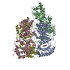
| Structure viewer | EM map: SurfViewMolmilJmol/JSmol |
| Supplemental images |

- Downloads & links
-EMDB archive
| Map data | emd_9380.map.gz | 4.6 MB | EMDB map data format | |
|---|---|---|---|---|
| Header (meta data) | emd-9380-v30.xmlemd-9380.xml | 14.2 KB 14.2 KB | Display Display | EMDB header |
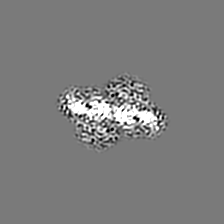
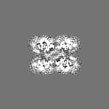
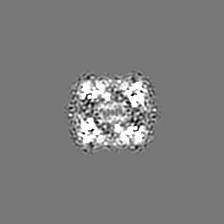
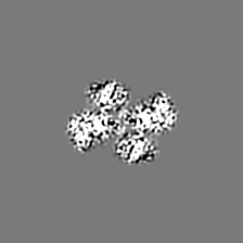
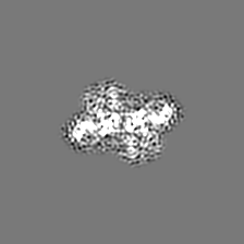
| Images | 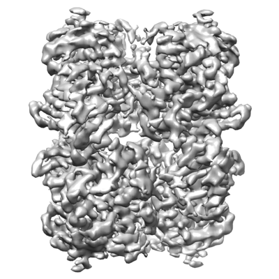 emd_9380.png emd_9380.png | 135.3 KB | ||
| Filedesc metadata | emd-9380.cif.gz | 6.1 KB | ||
| Archive directory |  http://ftp.pdbj.org/pub/emdb/structures/EMD-9380ftp://ftp.pdbj.org/pub/emdb/structures/EMD-9380 http://ftp.pdbj.org/pub/emdb/structures/EMD-9380ftp://ftp.pdbj.org/pub/emdb/structures/EMD-9380 | HTTPS FTP |
-Related structure data
| Related structure data | 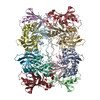 6nilMC 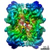 9381C M: atomic model generated by this map C: citing same article ( |
|---|---|
| Similar structure data |
-Links
| EMDB pages | EMDB (EBI/PDBe) / EMDataResource |
|---|---|
| Related items in Molecule of the Month |


-Map
| File | Download / File: emd_9380.map.gz / Format: CCP4 / Size: 42.9 MB / Type: IMAGE STORED AS FLOATING POINT NUMBER (4 BYTES) | ||||||||||||||||||||||||||||||||||||||||||||||||||||||||||||||||||||
|---|---|---|---|---|---|---|---|---|---|---|---|---|---|---|---|---|---|---|---|---|---|---|---|---|---|---|---|---|---|---|---|---|---|---|---|---|---|---|---|---|---|---|---|---|---|---|---|---|---|---|---|---|---|---|---|---|---|---|---|---|---|---|---|---|---|---|---|---|---|
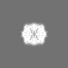
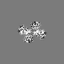
| Projections & slices | Image control
Images are generated by Spider. | ||||||||||||||||||||||||||||||||||||||||||||||||||||||||||||||||||||
| Voxel size | X=Y=Z: 1.05 Å | ||||||||||||||||||||||||||||||||||||||||||||||||||||||||||||||||||||
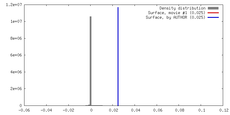
| Density |
| ||||||||||||||||||||||||||||||||||||||||||||||||||||||||||||||||||||
| Symmetry | Space group: 1 | ||||||||||||||||||||||||||||||||||||||||||||||||||||||||||||||||||||
| Details | EMDB XML:
CCP4 map header:
| ||||||||||||||||||||||||||||||||||||||||||||||||||||||||||||||||||||
 Z (Sec.)
 Y (Row.)
 X (Col.)


-Supplemental data
- Sample components
-Entire : truncated Vif/CBFbeta/A3Fctd complex
| Entire | Name: truncated Vif/CBFbeta/A3Fctd complex |
|---|---|
| Components |
|
-Supramolecule #1: truncated Vif/CBFbeta/A3Fctd complex
| Supramolecule | Name: truncated Vif/CBFbeta/A3Fctd complex / type: complex / ID: 1 / Parent: 0 / Macromolecule list: #1-#3 |
|---|---|
| Source (natural) | Organism: Human immunodeficiency virus 1 |
| Molecular weight | Theoretical: 17 KDa |
-Supramolecule #2: 6xHis tagged hA3Fctd-40aa-hCBFbeta fusion
| Supramolecule | Name: 6xHis tagged hA3Fctd-40aa-hCBFbeta fusion / type: complex / ID: 2 / Parent: 1 / Macromolecule list: #1-#2 Details: The N-terminus of human CBFbeta is fused to human A3Fctd through a 40 amino acid linker:GVDGSDEASELACPTPKEDGLAQQQTQLNLRSQATGSGSG |
|---|---|
| Source (natural) | Organism: Homo sapiens (human) |
-Supramolecule #3: alpha domain truncated HIV-1 Vif
| Supramolecule | Name: alpha domain truncated HIV-1 Vif / type: complex / ID: 3 / Parent: 1 / Macromolecule list: #3 |
|---|---|
| Source (natural) | Organism: Human immunodeficiency virus 1 |
-Macromolecule #1: DNA dC->dU-editing enzyme APOBEC-3F
| Macromolecule | Name: DNA dC->dU-editing enzyme APOBEC-3F / type: protein_or_peptide / ID: 1 / Number of copies: 4 / Enantiomer: LEVO / EC number: single-stranded DNA cytosine deaminase |
|---|---|
| Source (natural) | Organism: Homo sapiens (human) |
| Molecular weight | Theoretical: 24.433271 KDa |
| Recombinant expression | Organism:  |
| Sequence | String: MGSSHHHHHH SQDPNSMGKE ILRNPMEAMD PHIFYFHFKN LRKAYGRNES WLCFTMEVVK HHSPVSWKRG VFRNQVDPET GRHAERCFL SWFCDDILSP NTNYEVTWYT SWSPCPECAG EVAEFLARHS NVNLTIKTAR LYYFKDTDAA EGLRSLSQEG A SVEIMGYK ...String: MGSSHHHHHH SQDPNSMGKE ILRNPMEAMD PHIFYFHFKN LRKAYGRNES WLCFTMEVVK HHSPVSWKRG VFRNQVDPET GRHAERCFL SWFCDDILSP NTNYEVTWYT SWSPCPECAG EVAEFLARHS NVNLTIKTAR LYYFKDTDAA EGLRSLSQEG A SVEIMGYK DFKYCWENFV YNDDEPFKPW DGLDYNFLDL DSKLQEILE UniProtKB: DNA dC->dU-editing enzyme APOBEC-3F |
-Macromolecule #2: Core-binding factor subunit beta
| Macromolecule | Name: Core-binding factor subunit beta / type: protein_or_peptide / ID: 2 / Number of copies: 4 / Enantiomer: LEVO |
|---|---|
| Source (natural) | Organism: Homo sapiens (human) |
| Molecular weight | Theoretical: 17.851043 KDa |
| Recombinant expression | Organism: |
| Sequence | String: MPRVVPDQRS KFENEEFFRK LSRECEIKYT GFRDRPHEER QARFQNACRD GRSEIAFVAT GTNLSLQFFP ASWQGEQRQT PSREYVDLE REAGKVYLKA PMILNGVCVI WKGWIDLQRL DGMGCLEFDE ERAQQEDALA QQAFEEARRR TR UniProtKB: Core-binding factor subunit beta |
-Macromolecule #3: Virion infectivity factor
| Macromolecule | Name: Virion infectivity factor / type: protein_or_peptide / ID: 3 Details: Residues 114-157 was replaced with a 6 amino acid linker (EASEGS). The C-terminal residues 177-192 were deleted. Number of copies: 4 / Enantiomer: LEVO |
|---|---|
| Source (natural) | Organism: Human immunodeficiency virus 1 |
| Molecular weight | Theoretical: 16.736102 KDa |
| Recombinant expression | Organism: |
| Sequence | String: MENRWQVMIV WQVDRMRINT WKRLVKHHMY ISRKAKDWFY RHHYESTNPK ISSEVHIPLG DAKLVITTYW GLHTGERDWH LGQGVSIEW RKKRYSTQVD PDLADQLIHL HYFDEASEGS QIKPPLPSVR KLTEDRWNK UniProtKB: Virion infectivity factor, Virion infectivity factor |
-Macromolecule #4: ZINC ION
| Macromolecule | Name: ZINC ION / type: ligand / ID: 4 / Number of copies: 4 / Formula: ZN |
|---|---|
| Molecular weight | Theoretical: 65.409 Da |
-Experimental details
-Structure determination
| Method | cryo EM |
|---|---|
 Processing Processing | single particle reconstruction |
| Aggregation state | particle |
-Sample preparation
| Buffer | pH: 8 |
|---|---|
| Grid | Details: unspecified |
| Vitrification | Cryogen name: ETHANE |
- Electron microscopy
| Microscope | FEI TITAN KRIOS |
|---|---|
| Image recording | Film or detector model: GATAN K2 SUMMIT (4k x 4k) / Detector mode: SUPER-RESOLUTION / Average electron dose: 56.0 e/Å2 |
| Electron beam | Acceleration voltage: 300 kV / Electron source:  FIELD EMISSION GUN FIELD EMISSION GUN |
| Electron optics | C2 aperture diameter: 50.0 µm / Illumination mode: FLOOD BEAM / Imaging mode: BRIGHT FIELD / Cs: 2.7 mm |
| Experimental equipment |  Model: Titan Krios / Image courtesy: FEI Company |
-Image processing
| Startup model | Type of model: INSILICO MODEL |
|---|---|
| Final reconstruction | Applied symmetry - Point group: D2 (2x2 fold dihedral) / Resolution.type: BY AUTHOR / Resolution: 3.9 Å / Resolution method: FSC 0.143 CUT-OFF / Number images used: 337256 |
| Initial angle assignment | Type: PROJECTION MATCHING |
| Final angle assignment | Type: PROJECTION MATCHING |
