 Movie
Movie Controller


+ Open data

- Basic information
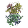
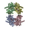
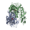
| Entry | Database: PDB / ID: 6nil | ||||||
|---|---|---|---|---|---|---|---|
| Title | cryoEM structure of the truncated HIV-1 Vif/CBFbeta/A3F complex | ||||||
 Components Components |
| ||||||
 Keywords Keywords | ANTIVIRAL PROTEIN / Human antiviral restriction factor / HIV viral protein | ||||||
| Function / homology |  Function and homology information Function and homology informationRUNX3 regulates RUNX1-mediated transcription / apolipoprotein B mRNA editing enzyme complex / RUNX1 regulates transcription of genes involved in BCR signaling / RUNX1 regulates transcription of genes involved in interleukin signaling / RUNX2 regulates bone development / core-binding factor complex / RUNX1 regulates expression of components of tight junctions / positive regulation of CD8-positive, alpha-beta T cell differentiation / RUNX2 regulates chondrocyte maturation / base conversion or substitution editing ...RUNX3 regulates RUNX1-mediated transcription / apolipoprotein B mRNA editing enzyme complex / RUNX1 regulates transcription of genes involved in BCR signaling / RUNX1 regulates transcription of genes involved in interleukin signaling / RUNX2 regulates bone development / core-binding factor complex / RUNX1 regulates expression of components of tight junctions / positive regulation of CD8-positive, alpha-beta T cell differentiation / RUNX2 regulates chondrocyte maturation / base conversion or substitution editing / single-stranded DNA cytosine deaminase / negative regulation of CD4-positive, alpha-beta T cell differentiation / negative regulation of single stranded viral RNA replication via double stranded DNA intermediate / DNA cytosine deamination / cytidine to uridine editing / clearance of foreign intracellular DNA / negative regulation of viral process / cytidine deaminase activity / RUNX1 and FOXP3 control the development of regulatory T lymphocytes (Tregs) / lymphocyte differentiation / RUNX2 regulates genes involved in cell migration / Transcriptional regulation by RUNX2 / RUNX1 regulates transcription of genes involved in differentiation of keratinocytes / RUNX2 regulates genes involved in differentiation of myeloid cells / positive regulation of gene expression via chromosomal CpG island demethylation / transposable element silencing / RUNX3 Regulates Immune Response and Cell Migration / myeloid cell differentiation / definitive hemopoiesis / RUNX1 regulates transcription of genes involved in differentiation of myeloid cells / Regulation of RUNX1 Expression and Activity / RUNX1 regulates transcription of genes involved in WNT signaling / RUNX1 regulates estrogen receptor mediated transcription / negative regulation of viral genome replication / RUNX2 regulates osteoblast differentiation / RUNX1 interacts with co-factors whose precise effect on RUNX1 targets is not known / RUNX3 regulates p14-ARF / viral life cycle / positive regulation of defense response to virus by host / cell maturation / antiviral innate immune response / P-body / RUNX1 regulates genes involved in megakaryocyte differentiation and platelet function / Regulation of RUNX3 expression and activity / virion component / Transcriptional regulation of granulopoiesis / protein polyubiquitination / osteoblast differentiation / Regulation of RUNX2 expression and activity / RUNX1 regulates transcription of genes involved in differentiation of HSCs / transcription by RNA polymerase II / Estrogen-dependent gene expression / sequence-specific DNA binding / defense response to virus / host cell cytoplasm / transcription coactivator activity / ribonucleoprotein complex / innate immune response / regulation of transcription by RNA polymerase II / host cell plasma membrane / negative regulation of transcription by RNA polymerase II / positive regulation of transcription by RNA polymerase II / RNA binding / zinc ion binding / nucleoplasm / membrane / identical protein binding / nucleus / cytoplasm Similarity search - Function | ||||||
| Biological species |  Homo sapiens (human) Homo sapiens (human)  Human immunodeficiency virus 1 Human immunodeficiency virus 1 | ||||||
| Method | ELECTRON MICROSCOPY / single particle reconstruction / cryo EM / Resolution: 3.9 Å | ||||||
 Authors Authors | Hu, Y. / Xiong, Y. | ||||||
| Funding support |  United States, 1items United States, 1items
| ||||||
 Citation Citation | Journal: Nat Struct Mol Biol / Year: 2019 Title: Structural basis of antagonism of human APOBEC3F by HIV-1 Vif. Authors: Yingxia Hu / Belete A Desimmie / Henry C Nguyen / Samantha J Ziegler / Tat Cheung Cheng / John Chen / Jia Wang / Hongwei Wang / Kai Zhang / Vinay K Pathak / Yong Xiong /   Abstract: HIV-1 virion infectivity factor (Vif) promotes degradation of the antiviral APOBEC3 (A3) proteins through the host ubiquitin-proteasome pathway to enable viral immune evasion. Disrupting Vif-A3 ...HIV-1 virion infectivity factor (Vif) promotes degradation of the antiviral APOBEC3 (A3) proteins through the host ubiquitin-proteasome pathway to enable viral immune evasion. Disrupting Vif-A3 interactions to reinstate the A3-catalyzed suppression of human immunodeficiency virus type 1 (HIV-1) replication is a potential approach for antiviral therapeutics. However, the molecular mechanisms by which Vif recognizes A3 proteins remain elusive. Here we report a cryo-EM structure of the Vif-targeted C-terminal domain of human A3F in complex with HIV-1 Vif and the cellular cofactor core-binding factor beta (CBFβ) at 3.9-Å resolution. The structure shows that Vif and CBFβ form a platform to recruit A3F, revealing a direct A3F-recruiting role of CBFβ beyond Vif stabilization, and captures multiple independent A3F-Vif interfaces. Together with our biochemical and cellular studies, our structural findings establish the molecular determinants that are critical for Vif-mediated neutralization of A3F and provide a comprehensive framework of how HIV-1 Vif hijacks the host protein degradation machinery to counteract viral restriction by A3F. | ||||||
| History |
|
- Structure visualization
| Movie |
Movie viewer |
|---|---|
| Structure viewer | Molecule: MolmilJmol/JSmol |
- Downloads & links
-Download
| PDBx/mmCIF format | 6nil.cif.gz | 383.8 KB | Display | PDBx/mmCIF format |
|---|---|---|---|---|
| PDB format | pdb6nil.ent.gz | 315 KB | Display | PDB format |
| PDBx/mmJSON format | 6nil.json.gz | Tree view | PDBx/mmJSON format | |
| Others |  Other downloads Other downloads |
-Validation report
| Arichive directory | https://data.pdbj.org/pub/pdb/validation_reports/ni/6nilftp://data.pdbj.org/pub/pdb/validation_reports/ni/6nil | HTTPS FTP |
|---|
-Related structure data
| Related structure data | 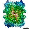 9380MC 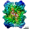 9381C M: map data used to model this data C: citing same article ( |
|---|---|
| Similar structure data |


-Links
 PDBj
PDBj


- Assembly
| Deposited unit | 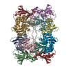
|
|---|---|
| 1 |
|
-Components
| #1: Protein | Mass: 24433.271 Da / Num. of mol.: 4 / Fragment: C-terminal domain Mutation: Y196D, H247G, C248R, F302K, W310K, Y314A, Q315A, K355D, K358D, F363D Source method: isolated from a genetically manipulated source Source: (gene. exp.) Homo sapiens (human) / Gene: APOBEC3F / Production host:  References: UniProt: Q8IUX4, single-stranded DNA cytosine deaminase #2: Protein | Mass: 17851.043 Da / Num. of mol.: 4 Source method: isolated from a genetically manipulated source Source: (gene. exp.) Homo sapiens (human) / Gene: CBFB / Production host: #3: Protein | Mass: 16736.102 Da / Num. of mol.: 4 Source method: isolated from a genetically manipulated source Details: Residues 114-157 was replaced with a 6 amino acid linker (EASEGS). The C-terminal residues 177-192 were deleted. Source: (gene. exp.) Human immunodeficiency virus 1 / Gene: vif / Production host: #4: Chemical | ChemComp-ZN /   Mass: 65.409 Da / Num. of mol.: 4 / Source method: obtained synthetically / Formula: Zn / Feature type: SUBJECT OF INVESTIGATION Mass: 65.409 Da / Num. of mol.: 4 / Source method: obtained synthetically / Formula: Zn / Feature type: SUBJECT OF INVESTIGATIONHas ligand of interest | Y | |
|---|
-Experimental details
-Experiment
| Experiment | Method: ELECTRON MICROSCOPY |
|---|---|
| EM experiment | Aggregation state: PARTICLE / 3D reconstruction method: single particle reconstruction |
- Sample preparation
| Component |
| ||||||||||||||||||||||||||||
|---|---|---|---|---|---|---|---|---|---|---|---|---|---|---|---|---|---|---|---|---|---|---|---|---|---|---|---|---|---|
| Molecular weight |
| ||||||||||||||||||||||||||||
| Source (natural) |
| ||||||||||||||||||||||||||||
| Source (recombinant) |
| ||||||||||||||||||||||||||||
| Buffer solution | pH: 8 | ||||||||||||||||||||||||||||
| Specimen | Embedding applied: NO / Shadowing applied: NO / Staining applied: NO / Vitrification applied: YES | ||||||||||||||||||||||||||||
| Specimen support | Details: unspecified | ||||||||||||||||||||||||||||
| Vitrification | Cryogen name: ETHANE |
- Electron microscopy imaging
| Experimental equipment |  Model: Titan Krios / Image courtesy: FEI Company |
|---|---|
| Microscopy | Model: FEI TITAN KRIOS |
| Electron gun | Electron source:  FIELD EMISSION GUN / Accelerating voltage: 300 kV / Illumination mode: FLOOD BEAM FIELD EMISSION GUN / Accelerating voltage: 300 kV / Illumination mode: FLOOD BEAM |
| Electron lens | Mode: BRIGHT FIELD / Cs: 2.7 mm / C2 aperture diameter: 50 µm |
| Image recording | Electron dose: 56 e/Å2 / Detector mode: SUPER-RESOLUTION / Film or detector model: GATAN K2 SUMMIT (4k x 4k) |
- Processing
| Software | Name: REFMAC / Version: 5.8.0257 / Classification: refinement |
|---|---|
| CTF correction | Type: NONE |
| Symmetry | Point symmetry: D2 (2x2 fold dihedral) |
| 3D reconstruction | Resolution: 3.9 Å / Resolution method: FSC 0.143 CUT-OFF / Num. of particles: 337256 / Symmetry type: POINT |
