 Movie
Movie Controller

[English] 日本語
 Yorodumi
- EMDB-9048: Ebola virus glycoprotein in complex with antigen binding fragment... -
+ Open data

- Basic information
| Entry | Database: EMDB / ID: EMD-9048 | |||||||||
|---|---|---|---|---|---|---|---|---|---|---|
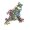
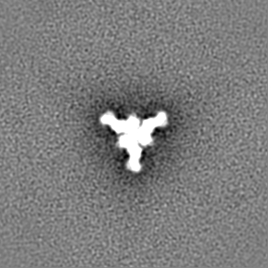
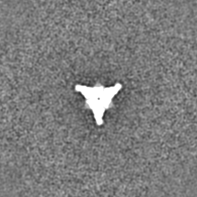
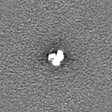
| Title | Ebola virus glycoprotein in complex with antigen binding fragment of chimeric mouse IgG 6D6 | |||||||||
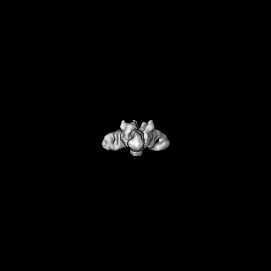
 Map data Map data | Ebola virus glycoprotein in complex with the antigen binding fragment of chimeric mouse IgG 6D6 | |||||||||
 Sample Sample |
| |||||||||
| Biological species |   Ebola virus - Mayinga, Zaire, 1976 / Ebola virus - Mayinga, Zaire, 1976 /  | |||||||||
| Method | single particle reconstruction / negative staining / Resolution: 20.0 Å | |||||||||
 Authors Authors | Milligan JC / Saphire EO | |||||||||
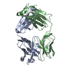
 Citation Citation | Journal: J Infect Dis / Year: 2019 Title: Structural Characterization of Pan-Ebolavirus Antibody 6D6 Targeting the Fusion Peptide of the Surface Glycoprotein. Authors: Jacob C Milligan / Diptiben V Parekh / Katherine M Fuller / Manabu Igarashi / Ayato Takada / Erica Ollmann Saphire /   Abstract: Ebola virus infection causes severe disease in humans and represents a global health threat. Candidates for immunotherapeutics and vaccines have shown promise in clinical trials, although they are ...Ebola virus infection causes severe disease in humans and represents a global health threat. Candidates for immunotherapeutics and vaccines have shown promise in clinical trials, although they are ineffective against other members of the Ebolavirus genus that also cause periodic, lethal outbreaks. In this study, we present a crystal structure of a pan-ebolavirus antibody, 6D6, as well as single-particle electron microscopy reconstructions of 6D6 in complex with Ebola and Bundibugyo virus glycoproteins. 6D6 binds to the conserved glycoprotein fusion peptide, implicating it as a site of immune vulnerability that could be exploited to reliably elicit a pan-ebolavirus neutralizing antibody response. | |||||||||
| History |
|
- Structure visualization
| Movie |
 Movie viewer Movie viewer |
|---|---|
| Structure viewer | EM map: SurfViewMolmilJmol/JSmol |
| Supplemental images |

- Downloads & links
-EMDB archive
| Map data | emd_9048.map.gz | 200.6 MB | EMDB map data format | |
|---|---|---|---|---|
| Header (meta data) | emd-9048-v30.xmlemd-9048.xml | 9.6 KB 9.6 KB | Display Display | EMDB header |
| Images | 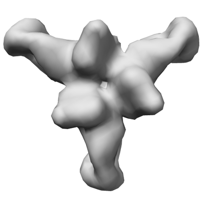 emd_9048.png emd_9048.png | 41.3 KB | ||
| Archive directory |  http://ftp.pdbj.org/pub/emdb/structures/EMD-9048ftp://ftp.pdbj.org/pub/emdb/structures/EMD-9048 http://ftp.pdbj.org/pub/emdb/structures/EMD-9048ftp://ftp.pdbj.org/pub/emdb/structures/EMD-9048 | HTTPS FTP |
-Validation report
| Summary document | emd_9048_validation.pdf.gz | 77.9 KB | Display | EMDB validaton report |
|---|---|---|---|---|
| Full document | emd_9048_full_validation.pdf.gz | 77 KB | Display | |
| Data in XML | emd_9048_validation.xml.gz | 494 B | Display | |
| Arichive directory | https://ftp.pdbj.org/pub/emdb/validation_reports/EMD-9048ftp://ftp.pdbj.org/pub/emdb/validation_reports/EMD-9048 | HTTPS FTP |
-Related structure data


-Links
| EMDB pages | EMDB (EBI/PDBe) / EMDataResource |
|---|
-Map
| File | Download / File: emd_9048.map.gz / Format: CCP4 / Size: 216 MB / Type: IMAGE STORED AS FLOATING POINT NUMBER (4 BYTES) | ||||||||||||||||||||||||||||||||||||||||||||||||||||||||||||
|---|---|---|---|---|---|---|---|---|---|---|---|---|---|---|---|---|---|---|---|---|---|---|---|---|---|---|---|---|---|---|---|---|---|---|---|---|---|---|---|---|---|---|---|---|---|---|---|---|---|---|---|---|---|---|---|---|---|---|---|---|---|
| Annotation | Ebola virus glycoprotein in complex with the antigen binding fragment of chimeric mouse IgG 6D6 | ||||||||||||||||||||||||||||||||||||||||||||||||||||||||||||
| Projections & slices | Image control
Images are generated by Spider. | ||||||||||||||||||||||||||||||||||||||||||||||||||||||||||||
| Voxel size | X=Y=Z: 1.66 Å | ||||||||||||||||||||||||||||||||||||||||||||||||||||||||||||
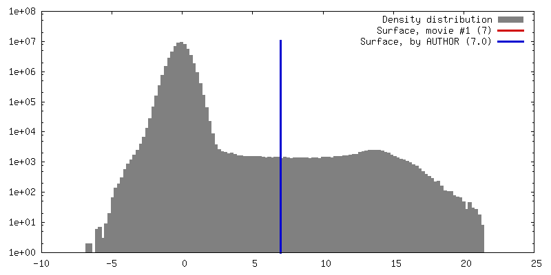
| Density |
| ||||||||||||||||||||||||||||||||||||||||||||||||||||||||||||
| Symmetry | Space group: 1 | ||||||||||||||||||||||||||||||||||||||||||||||||||||||||||||
| Details | EMDB XML:
CCP4 map header:
| ||||||||||||||||||||||||||||||||||||||||||||||||||||||||||||
 Z (Sec.)
 Y (Row.)
 X (Col.)


-Supplemental data
- Sample components
-Entire : Ebola virus glycoprotein in complex with antigen binding fragment...
| Entire | Name: Ebola virus glycoprotein in complex with antigen binding fragment of chimeric mouse IgG 6D6 |
|---|---|
| Components |
|
-Supramolecule #1: Ebola virus glycoprotein in complex with antigen binding fragment...
| Supramolecule | Name: Ebola virus glycoprotein in complex with antigen binding fragment of chimeric mouse IgG 6D6 type: complex / ID: 1 / Parent: 0 |
|---|---|
| Source (natural) | Organism: Ebola virus - Mayinga, Zaire, 1976 |
| Recombinant expression | Organism:  |
| Molecular weight | Theoretical: 150 KDa |
-Supramolecule #2: Antigen binding fragment of chimeric mouse IgG 6D6
| Supramolecule | Name: Antigen binding fragment of chimeric mouse IgG 6D6 / type: complex / ID: 2 / Parent: 1 |
|---|---|
| Source (natural) | Organism: |
| Recombinant expression | Organism: |
-Supramolecule #3: Ebola virus glycoprotein
| Supramolecule | Name: Ebola virus glycoprotein / type: complex / ID: 3 / Parent: 1 |
|---|---|
| Source (natural) | Organism: Ebola virus - Mayinga, Zaire, 1976 |
| Recombinant expression | Organism: |
-Experimental details
-Structure determination
| Method | negative staining |
|---|---|
 Processing Processing | single particle reconstruction |
| Aggregation state | particle |
-Sample preparation
| Concentration | 0.01 mg/mL | ||||||
|---|---|---|---|---|---|---|---|
| Buffer | pH: 7.5 / Component:
| ||||||
| Staining | Type: NEGATIVE / Material: Uranyl formate | ||||||
| Grid | Material: COPPER / Support film - Material: CARBON / Support film - topology: CONTINUOUS |
- Electron microscopy
| Microscope | FEI TITAN |
|---|---|
| Image recording | Film or detector model: FEI FALCON II (4k x 4k) / Average electron dose: 40.0 e/Å2 |
| Electron beam | Acceleration voltage: 300 kV / Electron source:  FIELD EMISSION GUN FIELD EMISSION GUN |
| Electron optics | Illumination mode: FLOOD BEAM / Imaging mode: BRIGHT FIELD |
-Image processing
| Final reconstruction | Resolution.type: BY AUTHOR / Resolution: 20.0 Å / Resolution method: OTHER / Details: Negative stain resolution / Number images used: 6187 |
|---|---|
| Initial angle assignment | Type: MAXIMUM LIKELIHOOD |
| Final angle assignment | Type: MAXIMUM LIKELIHOOD |
-Atomic model buiding 1
| Refinement | Protocol: AB INITIO MODEL |
|---|
