 Movie
Movie Controller

[English] 日本語
 Yorodumi
- EMDB-8371: Three-dimensional reconstruction of bat adenovirus C isolate 250-A -
+ Open data

- Basic information
| Entry | Database: EMDB / ID: EMD-8371 | |||||||||
|---|---|---|---|---|---|---|---|---|---|---|
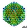
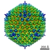
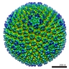
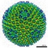
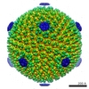
| Title | Three-dimensional reconstruction of bat adenovirus C isolate 250-A | |||||||||
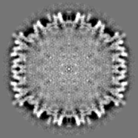
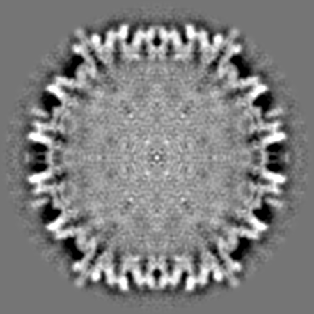
 Map data Map data | The bat adenovirus isolate 250-A reconstruction by cryo-EM | |||||||||
 Sample Sample |
| |||||||||
| Biological species |  Bat adenovirus Bat adenovirus | |||||||||
| Method | single particle reconstruction / cryo EM / Resolution: 17.9 Å | |||||||||
 Authors Authors | Hackenbrack NM / Hafenstein SL | |||||||||
 Citation Citation | Journal: Science / Year: 2010 Title: Atomic structure of human adenovirus by cryo-EM reveals interactions among protein networks. Authors: Hongrong Liu / Lei Jin / Sok Boon S Koh / Ivo Atanasov / Stan Schein / Lily Wu / Z Hong Zhou /  Abstract: Construction of a complex virus may involve a hierarchy of assembly elements. Here, we report the structure of the whole human adenovirus virion at 3.6 angstroms resolution by cryo-electron ...Construction of a complex virus may involve a hierarchy of assembly elements. Here, we report the structure of the whole human adenovirus virion at 3.6 angstroms resolution by cryo-electron microscopy (cryo-EM), revealing in situ atomic models of three minor capsid proteins (IIIa, VIII, and IX), extensions of the (penton base and hexon) major capsid proteins, and interactions within three protein-protein networks. One network is mediated by protein IIIa at the vertices, within group-of-six (GOS) tiles--a penton base and its five surrounding hexons. Another is mediated by ropes (protein IX) that lash hexons together to form group-of-nine (GON) tiles and bind GONs to GONs. The third, mediated by IIIa and VIII, binds each GOS to five surrounding GONs. Optimization of adenovirus for cancer and gene therapy could target these networks. | |||||||||
| History |
|
- Structure visualization
| Movie |
 Movie viewer Movie viewer |
|---|---|
| Structure viewer | EM map: SurfViewMolmilJmol/JSmol |
| Supplemental images |
 UCSF Chimera
- Downloads & links
-EMDB archive
| Map data | emd_8371.map.gz | 146.8 MB | EMDB map data format | |
|---|---|---|---|---|
| Header (meta data) | emd-8371-v30.xmlemd-8371.xml | 13.9 KB 13.9 KB | Display Display | EMDB header |
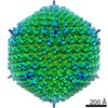
| Images |  emd_8371.png emd_8371.png | 268 KB | ||
| Archive directory |  http://ftp.pdbj.org/pub/emdb/structures/EMD-8371ftp://ftp.pdbj.org/pub/emdb/structures/EMD-8371 http://ftp.pdbj.org/pub/emdb/structures/EMD-8371ftp://ftp.pdbj.org/pub/emdb/structures/EMD-8371 | HTTPS FTP |
-Validation report
| Summary document | emd_8371_validation.pdf.gz | 78 KB | Display | EMDB validaton report |
|---|---|---|---|---|
| Full document | emd_8371_full_validation.pdf.gz | 77.1 KB | Display | |
| Data in XML | emd_8371_validation.xml.gz | 494 B | Display | |
| Arichive directory | https://ftp.pdbj.org/pub/emdb/validation_reports/EMD-8371ftp://ftp.pdbj.org/pub/emdb/validation_reports/EMD-8371 | HTTPS FTP |
-Related structure data
| Related structure data |  5172 |
|---|---|
| Similar structure data |


-Links
| EMDB pages | EMDB (EBI/PDBe) / EMDataResource |
|---|
-Map
| File | Download / File: emd_8371.map.gz / Format: CCP4 / Size: 349.9 MB / Type: IMAGE STORED AS FLOATING POINT NUMBER (4 BYTES) | ||||||||||||||||||||||||||||||||||||||||||||||||||||||||||||||||||||
|---|---|---|---|---|---|---|---|---|---|---|---|---|---|---|---|---|---|---|---|---|---|---|---|---|---|---|---|---|---|---|---|---|---|---|---|---|---|---|---|---|---|---|---|---|---|---|---|---|---|---|---|---|---|---|---|---|---|---|---|---|---|---|---|---|---|---|---|---|---|
| Annotation | The bat adenovirus isolate 250-A reconstruction by cryo-EM | ||||||||||||||||||||||||||||||||||||||||||||||||||||||||||||||||||||
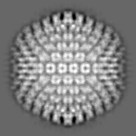
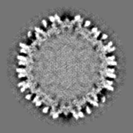
| Projections & slices | Image control
Images are generated by Spider. | ||||||||||||||||||||||||||||||||||||||||||||||||||||||||||||||||||||
| Voxel size | X=Y=Z: 2.33 Å | ||||||||||||||||||||||||||||||||||||||||||||||||||||||||||||||||||||
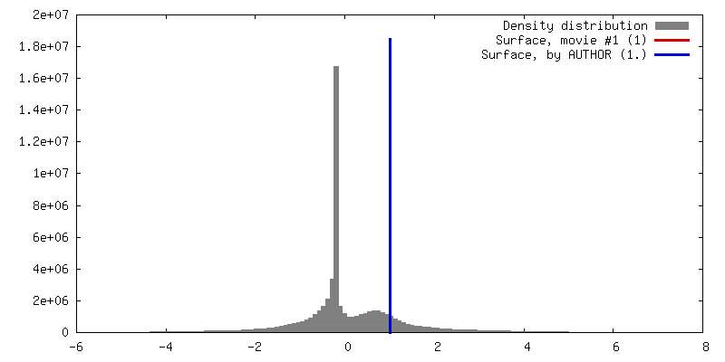
| Density |
| ||||||||||||||||||||||||||||||||||||||||||||||||||||||||||||||||||||
| Symmetry | Space group: 1 | ||||||||||||||||||||||||||||||||||||||||||||||||||||||||||||||||||||
| Details | EMDB XML:
CCP4 map header:
| ||||||||||||||||||||||||||||||||||||||||||||||||||||||||||||||||||||
 Z (Sec.)
 Y (Row.)
 X (Col.)


-Supplemental data
- Sample components
-Entire : Bat adenovirus
| Entire | Name: Bat adenovirus |
|---|---|
| Components |
|
-Supramolecule #1: Bat adenovirus
| Supramolecule | Name: Bat adenovirus / type: virus / ID: 1 / Parent: 0 Details: Bat adenovirus C isolate 250-A was isolated from Corynorhinus rafinesquii at the Mammoth Cave National Park in Kentucky, USA NCBI-ID: 740971 / Sci species name: Bat adenovirus / Sci species strain: C / Virus type: VIRION / Virus isolate: SPECIES / Virus enveloped: No / Virus empty: No |
|---|---|
| Host (natural) | Organism:  Plecotus rafinesquii (Rafinesque's big-eared bat) Plecotus rafinesquii (Rafinesque's big-eared bat) |
-Experimental details
-Structure determination
| Method | cryo EM |
|---|---|
 Processing Processing | single particle reconstruction |
| Aggregation state | cell |
-Sample preparation
| Buffer | pH: 7.5 Component:
| ||||||||||||
|---|---|---|---|---|---|---|---|---|---|---|---|---|---|
| Grid | Model: Quantifoil / Material: COPPER / Support film - Material: CARBON / Support film - topology: CONTINUOUS / Support film - Film thickness: 7.0 nm / Pretreatment - Type: GLOW DISCHARGE / Pretreatment - Atmosphere: OTHER / Pretreatment - Pressure: 2e-06 kPa | ||||||||||||
| Vitrification | Cryogen name: ETHANE / Chamber humidity: 95 % / Instrument: GATAN CRYOPLUNGE 3 | ||||||||||||
| Details | Tissue from Corynorhinus rafinesquii was homogenized, clarified, and then used to infect Vero E6 cells. The supernatant was harvested and spun on a cesium chloride step gradient. Full particles were harvested, dialyzed, and concentrated. |
- Electron microscopy
| Microscope | JEOL 2100 |
|---|---|
| Image recording | Film or detector model: GATAN ULTRASCAN 4000 (4k x 4k) / Number real images: 383 / Average electron dose: 10.0 e/Å2 |
| Electron beam | Acceleration voltage: 200 kV / Electron source: LAB6 |
| Electron optics | Illumination mode: OTHER / Imaging mode: OTHER |
| Sample stage | Specimen holder model: GATAN 626 SINGLE TILT LIQUID NITROGEN CRYO TRANSFER HOLDER |
