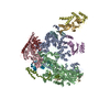
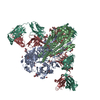
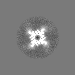
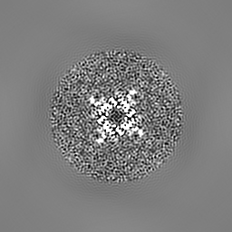
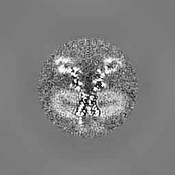
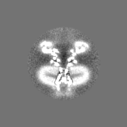
登録情報 データベース : EMDB / ID : EMD-8215タイトル Single particle cryo-EM structure of the voltage-gated K+ channel Eag1 bound to the channel inhibitor calmodulin Voltage-gated K+ channel Eag1 bound to the channel inhibitor calmodulin 細胞器官・細胞要素 : Voltage-gated K+ channel Eag1 bound to the channel inhibitor calmodulin細胞器官・細胞要素 : Voltage-gated potassium channel Eag1タンパク質・ペプチド : Potassium voltage-gated channel subfamily H member 1細胞器官・細胞要素 : Calmodulinリガンド : 2-acetamido-2-deoxy-beta-D-glucopyranoseリガンド : CHOLESTEROL HEMISUCCINATE / / / / 機能・相同性 分子機能 ドメイン・相同性 構成要素
/ / / / / / / / / / / / / / / / / / / / / / / / / / / / / / / / / / / / / / / / / / / / / / / / / / / / / / / / / / / / / / / / / / / / / / / / / / / / / / / / / / / / / / / / / / / / / / / / / / / / / / / / / / / / / / / / / / / / / / / / / / / / / / / / / / / / / / / / / / / / / / / / / 生物種 Rattus norvegicus (ドブネズミ) / Homo sapiens (ヒト)手法 / / 解像度 : 3.78 Å Whicher JR / MacKinnon R 資金援助 Organization Grant number 国 National Institutes of Health/National Institute of General Medical Sciences (NIH/NIGMS) GM43949 Howard Hughes Medical Institute (HHMI) Damon Runyon Cancer Research Foundation DRG-2212-15
ジャーナル : Science / 年 : 2016タイトル : Structure of the voltage-gated K⁺ channel Eag1 reveals an alternative voltage sensing mechanism.著者 : Jonathan R Whicher / Roderick MacKinnon / 要旨 : Voltage-gated potassium (K(v)) channels are gated by the movement of the transmembrane voltage sensor, which is coupled, through the helical S4-S5 linker, to the potassium pore. We determined the ... Voltage-gated potassium (K(v)) channels are gated by the movement of the transmembrane voltage sensor, which is coupled, through the helical S4-S5 linker, to the potassium pore. We determined the single-particle cryo-electron microscopy structure of mammalian K(v)10.1, or Eag1, bound to the channel inhibitor calmodulin, at 3.78 angstrom resolution. Unlike previous K(v) structures, the S4-S5 linker of Eag1 is a five-residue loop and the transmembrane segments are not domain swapped, which suggest an alternative mechanism of voltage-dependent gating. Additionally, the structure and position of the S4-S5 linker allow calmodulin to bind to the intracellular domains and to close the potassium pore, independent of voltage-sensor position. The structure reveals an alternative gating mechanism for K(v) channels and provides a template to further understand the gating properties of Eag1 and related channels. 履歴 登録 2016年6月16日 - ヘッダ(付随情報) 公開 2016年8月17日 - マップ公開 2016年8月17日 - 更新 2024年10月23日 - 現状 2024年10月23日 処理サイト : RCSB / 状態 : 公開
すべて表示 表示を減らす
 ムービー
ムービー コントローラー


 データを開く
 基本情報
基本情報 マップデータ
 試料
試料 キーワード
キーワード 機能・相同性情報
 Homo sapiens (ヒト)
 データ登録者
 米国, 3件
  引用
引用 構造の表示
構造の表示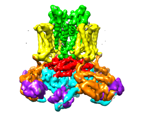
 ダウンロードとリンク
 emd_8215.png
 http://ftp.pdbj.org/pub/emdb/structures/EMD-8215


 Z (Sec.)
 Y (Row.)
 X (Col.)


 試料の構成要素

 解析
解析 電子顕微鏡法
 FIELD EMISSION GUN


