Movie
Movie Controller

[English] 日本語
 Yorodumi
- EMDB-20294: Single particle cryo-EM structure of the voltage-gated K+ channel... -
+ Open data

- Basic information
| Entry | Database: EMDB / ID: EMD-20294 | ||||||||||||
|---|---|---|---|---|---|---|---|---|---|---|---|---|---|
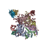
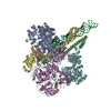
| Title | Single particle cryo-EM structure of the voltage-gated K+ channel Eag1 3-13 deletion mutant bound to calmodulin (conformation 2) | ||||||||||||
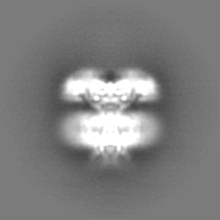
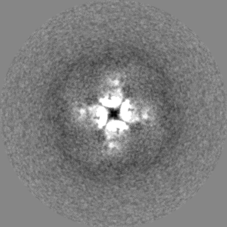
 Map data Map data | Sharpened map for Eag1 3-13/CaM conformation 2 | ||||||||||||
 Sample Sample |
| ||||||||||||
 Keywords Keywords | Voltage-gated potassium channel / ion channel / calmodulin / TRANSPORT PROTEIN-CALCIUM BINDING PROTEIN complex | ||||||||||||
| Function / homology |  Function and homology information Function and homology informationVoltage gated Potassium channels / potassium channel complex / regulation of presynaptic cytosolic calcium ion concentration / voltage-gated monoatomic ion channel activity involved in regulation of presynaptic membrane potential / delayed rectifier potassium channel activity / nuclear inner membrane / CaM pathway / Cam-PDE 1 activation / Sodium/Calcium exchangers / Calmodulin induced events ...Voltage gated Potassium channels / potassium channel complex / regulation of presynaptic cytosolic calcium ion concentration / voltage-gated monoatomic ion channel activity involved in regulation of presynaptic membrane potential / delayed rectifier potassium channel activity / nuclear inner membrane / CaM pathway / Cam-PDE 1 activation / Sodium/Calcium exchangers / Calmodulin induced events / Reduction of cytosolic Ca++ levels / Activation of Ca-permeable Kainate Receptor / CREB1 phosphorylation through the activation of CaMKII/CaMKK/CaMKIV cascasde / Loss of phosphorylation of MECP2 at T308 / CREB1 phosphorylation through the activation of Adenylate Cyclase / negative regulation of high voltage-gated calcium channel activity / PKA activation / CaMK IV-mediated phosphorylation of CREB / phosphatidylinositol bisphosphate binding / Glycogen breakdown (glycogenolysis) / negative regulation of ryanodine-sensitive calcium-release channel activity / Activation of RAC1 downstream of NMDARs / organelle localization by membrane tethering / CLEC7A (Dectin-1) induces NFAT activation / : / autophagosome membrane docking / regulation of synaptic vesicle exocytosis / negative regulation of calcium ion export across plasma membrane / regulation of cardiac muscle cell action potential / presynaptic endocytosis / startle response / Synthesis of IP3 and IP4 in the cytosol / Phase 0 - rapid depolarisation / Negative regulation of NMDA receptor-mediated neuronal transmission / parallel fiber to Purkinje cell synapse / Unblocking of NMDA receptors, glutamate binding and activation / calcineurin-mediated signaling / RHO GTPases activate PAKs / regulation of cell communication by electrical coupling involved in cardiac conduction / Ion transport by P-type ATPases / Uptake and function of anthrax toxins / protein phosphatase activator activity / regulation of ryanodine-sensitive calcium-release channel activity / Long-term potentiation / Calcineurin activates NFAT / Regulation of MECP2 expression and activity / DARPP-32 events / Smooth Muscle Contraction / axolemma / detection of calcium ion / catalytic complex / regulation of cardiac muscle contraction / cellular response to interferon-beta / RHO GTPases activate IQGAPs / calcium channel inhibitor activity / presynaptic cytosol / Activation of AMPK downstream of NMDARs / regulation of release of sequestered calcium ion into cytosol by sarcoplasmic reticulum / eNOS activation / Ion homeostasis / Tetrahydrobiopterin (BH4) synthesis, recycling, salvage and regulation / regulation of calcium-mediated signaling / Protein methylation / titin binding / regulation of cardiac muscle contraction by regulation of the release of sequestered calcium ion / 14-3-3 protein binding / voltage-gated potassium channel complex / FCERI mediated Ca+2 mobilization / calcium channel complex / substantia nigra development / potassium ion transmembrane transport / regulation of heart rate / FCGR3A-mediated IL10 synthesis / cellular response to calcium ion / calyx of Held / Ras activation upon Ca2+ influx through NMDA receptor / Antigen activates B Cell Receptor (BCR) leading to generation of second messengers / adenylate cyclase activator activity / potassium ion transport / protein serine/threonine kinase activator activity / VEGFR2 mediated cell proliferation / VEGFR2 mediated vascular permeability / regulation of cytokinesis / spindle microtubule / positive regulation of receptor signaling pathway via JAK-STAT / sarcomere / Translocation of SLC2A4 (GLUT4) to the plasma membrane / calcium channel regulator activity / regulation of membrane potential / Transcriptional activation of mitochondrial biogenesis / RAF activation / response to calcium ion / cellular response to type II interferon / postsynaptic density membrane / G2/M transition of mitotic cell cycle / Stimuli-sensing channels / long-term synaptic potentiation / spindle pole / calcium-dependent protein binding / Signaling by RAF1 mutants Similarity search - Function | ||||||||||||
| Biological species |   Homo sapiens (human) Homo sapiens (human) | ||||||||||||
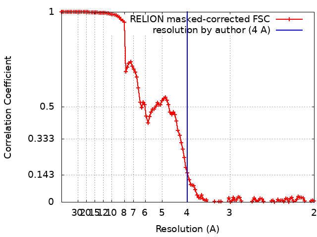
| Method | single particle reconstruction / cryo EM / Resolution: 4.0 Å | ||||||||||||
 Authors Authors | Whicher JR / MacKinnon R | ||||||||||||
| Funding support |  United States, 3 items United States, 3 items
| ||||||||||||
 Citation Citation | Journal: Elife / Year: 2019 Title: Regulation of Eag1 gating by its intracellular domains. Authors: Jonathan R Whicher / Roderick MacKinnon / Abstract: Voltage-gated potassium channels (Ks) are gated by transmembrane voltage sensors (VS) that move in response to changes in membrane voltage. K10.1 or Eag1 also has three intracellular domains: PAS, C- ...Voltage-gated potassium channels (Ks) are gated by transmembrane voltage sensors (VS) that move in response to changes in membrane voltage. K10.1 or Eag1 also has three intracellular domains: PAS, C-linker, and CNBHD. We demonstrate that the Eag1 intracellular domains are not required for voltage-dependent gating but likely interact with the VS to modulate gating. We identified specific interactions between the PAS, CNBHD, and VS that modulate voltage-dependent gating and provide evidence that VS movement destabilizes these interactions to promote channel opening. Additionally, mutation of these interactions renders Eag1 insensitive to calmodulin inhibition. The structure of the calmodulin insensitive mutant in a pre-open conformation suggests that channel opening may occur through a rotation of the intracellular domains and calmodulin may prevent this rotation by stabilizing interactions between the VS and intracellular domains. Intracellular domains likely play a similar modulatory role in voltage-dependent gating of the related K11-12 channels. | ||||||||||||
| History |
|
- Structure visualization
| Movie |
Movie viewer |
|---|---|
| Structure viewer | EM map: SurfViewMolmilJmol/JSmol |
| Supplemental images |

- Downloads & links
-EMDB archive
| Map data | emd_20294.map.gz | 116.9 MB | EMDB map data format | |
|---|---|---|---|---|
| Header (meta data) | emd-20294-v30.xmlemd-20294.xml | 16 KB 16 KB | Display Display | EMDB header |
| FSC (resolution estimation) | emd_20294_fsc.xml | 11.2 KB | Display | FSC data file |
| Images | 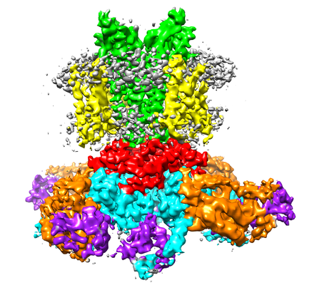 emd_20294.png emd_20294.png | 196.3 KB | ||
| Filedesc metadata | emd-20294.cif.gz | 6.1 KB | ||
| Others | emd_20294_additional.map.gz | 92.9 MB | ||
| Archive directory |  http://ftp.pdbj.org/pub/emdb/structures/EMD-20294ftp://ftp.pdbj.org/pub/emdb/structures/EMD-20294 http://ftp.pdbj.org/pub/emdb/structures/EMD-20294ftp://ftp.pdbj.org/pub/emdb/structures/EMD-20294 | HTTPS FTP |
-Related structure data
| Related structure data |  6pbxMC  6pbyC C: citing same article ( M: atomic model generated by this map |
|---|---|
| Similar structure data |


-Links
| EMDB pages | EMDB (EBI/PDBe) / EMDataResource |
|---|---|
| Related items in Molecule of the Month |


-Map
| File | Download / File: emd_20294.map.gz / Format: CCP4 / Size: 125 MB / Type: IMAGE STORED AS FLOATING POINT NUMBER (4 BYTES) | ||||||||||||||||||||||||||||||||||||||||||||||||||||||||||||||||||||
|---|---|---|---|---|---|---|---|---|---|---|---|---|---|---|---|---|---|---|---|---|---|---|---|---|---|---|---|---|---|---|---|---|---|---|---|---|---|---|---|---|---|---|---|---|---|---|---|---|---|---|---|---|---|---|---|---|---|---|---|---|---|---|---|---|---|---|---|---|---|
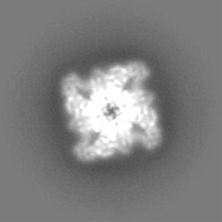
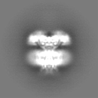
| Annotation | Sharpened map for Eag1 3-13/CaM conformation 2 | ||||||||||||||||||||||||||||||||||||||||||||||||||||||||||||||||||||
| Projections & slices | Image control
Images are generated by Spider. | ||||||||||||||||||||||||||||||||||||||||||||||||||||||||||||||||||||
| Voxel size | X=Y=Z: 1 Å | ||||||||||||||||||||||||||||||||||||||||||||||||||||||||||||||||||||
| Density |
| ||||||||||||||||||||||||||||||||||||||||||||||||||||||||||||||||||||
| Symmetry | Space group: 1 | ||||||||||||||||||||||||||||||||||||||||||||||||||||||||||||||||||||
| Details | EMDB XML:
CCP4 map header:
| ||||||||||||||||||||||||||||||||||||||||||||||||||||||||||||||||||||
 Z (Sec.)
 Y (Row.)
 X (Col.)


-Supplemental data
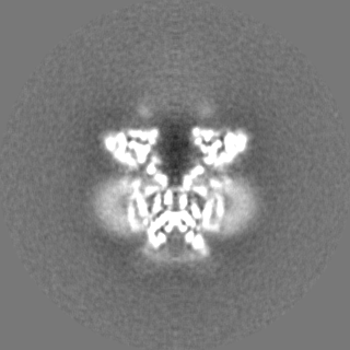
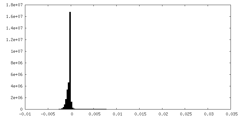
-Additional map: Unsharpened map for Eag1 3-13/CaM conformation 2
| File | emd_20294_additional.map | ||||||||||||
|---|---|---|---|---|---|---|---|---|---|---|---|---|---|
| Annotation | Unsharpened map for Eag1 3-13/CaM conformation 2 | ||||||||||||
| Projections & Slices |
| ||||||||||||
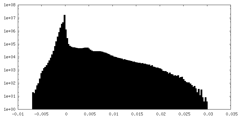
| Density Histograms |

- Sample components
-Entire : Voltage-gated potassium channel Eag1 3-13 deletion mutant bound t...
| Entire | Name: Voltage-gated potassium channel Eag1 3-13 deletion mutant bound to calmodulin |
|---|---|
| Components |
|
-Supramolecule #1: Voltage-gated potassium channel Eag1 3-13 deletion mutant bound t...
| Supramolecule | Name: Voltage-gated potassium channel Eag1 3-13 deletion mutant bound to calmodulin type: complex / ID: 1 / Parent: 0 / Macromolecule list: all |
|---|
-Supramolecule #2: Voltage-gated potassium channel Eag1 3-13 deletion mutant
| Supramolecule | Name: Voltage-gated potassium channel Eag1 3-13 deletion mutant type: complex / ID: 2 / Parent: 1 / Macromolecule list: #1 |
|---|---|
| Source (natural) | Organism: |
-Supramolecule #3: calmodulin
| Supramolecule | Name: calmodulin / type: complex / ID: 3 / Parent: 1 / Macromolecule list: #2 |
|---|---|
| Source (natural) | Organism: Homo sapiens (human) |
-Macromolecule #1: Potassium voltage-gated channel subfamily H member 1
| Macromolecule | Name: Potassium voltage-gated channel subfamily H member 1 / type: protein_or_peptide / ID: 1 / Number of copies: 4 / Enantiomer: LEVO |
|---|---|
| Source (natural) | Organism: |
| Molecular weight | Theoretical: 96.26143 KDa |
| Recombinant expression | Organism: Homo sapiens (human) |
| Sequence | String: MAQNTFLENI VRRSNDTNFV LGNAQIVDWP IVYSNDGFCK LSGYHRAEVM QKSSACSFMY GELTDKDTVE KVRQTFENYE MNSFEILMY KKNRTPVWFF VKIAPIRNEQ DKVVLFLCTF SDITAFKQPI EDDSCKGWGK FARLTRALTS SRGVLQQLAP S VQKGENVH ...String: MAQNTFLENI VRRSNDTNFV LGNAQIVDWP IVYSNDGFCK LSGYHRAEVM QKSSACSFMY GELTDKDTVE KVRQTFENYE MNSFEILMY KKNRTPVWFF VKIAPIRNEQ DKVVLFLCTF SDITAFKQPI EDDSCKGWGK FARLTRALTS SRGVLQQLAP S VQKGENVH KHSRLAEVLQ LGSDILPQYK QEAPKTPPHI ILHYCVFKTT WDWIILILTF YTAILVPYNV SFKTRQNNVA WL VVDSIVD VIFLVDIVLN FHTTFVGPAG EVISDPKLIR MNYLKTWFVI DLLSCLPYDV INAFENVDEG ISSLFSSLKV VRL LRLGRV ARKLDHYIEY GAAVLVLLVC VFGLAAHWMA CIWYSIGDYE IFDEDTKTIR NNSWLYQLAL DIGTPYQFNG SGSG KWEGG PSKNSVYISS LYFTMTSLTS VGFGNIAPST DIEKIFAVAI MMIGSLLYAT IFGNVTTIFQ QMYANTNRYH EMLNS VRDF LKLYQVPKGL SERVMDYIVS TWSMSRGIDT EKVLQICPKD MRADICVHLN RKVFKEHPAF RLASDGCLRA LAMEFQ TVH CAPGDLIYHA GESVDSLCFV VSGSLEVIQD DEVVAILGKG DVFGDVFWKE ATLAQSCANV RALTYCDLHV IKRDALQ KV LEFYTAFSHS FSRNLILTYN LRKRIVFRKI SDVKREEEER MKRKNEAPLI LPPDHPVRRL FQRFRQQKEA RLAAERGG R DLDDLDVEKG NALTDHTSAN HSLVKASVVT VRESPATPVS FYPIPEQTLQ ATVLEVKHEL KEDIKALNAK MTSIEKQLS EILRILMSRG SSQSPQDTCE VSRPQSPESD RDIFGASSNS LEVLFQ UniProtKB: Voltage-gated delayed rectifier potassium channel KCNH1, Voltage-gated delayed rectifier potassium channel KCNH1 |
-Macromolecule #2: Calmodulin-1
| Macromolecule | Name: Calmodulin-1 / type: protein_or_peptide / ID: 2 / Number of copies: 4 / Enantiomer: LEVO |
|---|---|
| Source (natural) | Organism: Homo sapiens (human) |
| Molecular weight | Theoretical: 16.852545 KDa |
| Recombinant expression | Organism: Homo sapiens (human) |
| Sequence | String: MADQLTEEQI AEFKEAFSLF DKDGDGTITT KELGTVMRSL GQNPTEAELQ DMINEVDADG NGTIDFPEFL TMMARKMKDT DSEEEIREA FRVFDKDGNG YISAAELRHV MTNLGEKLTD EEVDEMIREA DIDGDGQVNY EEFVQMMTAK UniProtKB: Calmodulin-1 |
-Experimental details
-Structure determination
| Method | cryo EM |
|---|---|
 Processing Processing | single particle reconstruction |
| Aggregation state | particle |
-Sample preparation
| Buffer | pH: 8 |
|---|---|
| Grid | Details: unspecified |
| Vitrification | Cryogen name: ETHANE |
- Electron microscopy
| Microscope | FEI TITAN KRIOS |
|---|---|
| Image recording | Film or detector model: GATAN K2 SUMMIT (4k x 4k) / Average electron dose: 1.6 e/Å2 |
| Electron beam | Acceleration voltage: 300 kV / Electron source:  FIELD EMISSION GUN FIELD EMISSION GUN |
| Electron optics | Illumination mode: FLOOD BEAM / Imaging mode: BRIGHT FIELD |
| Experimental equipment |  Model: Titan Krios / Image courtesy: FEI Company |