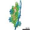
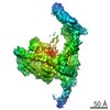
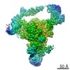
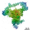
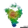
ジャーナル: Nature / 年: 2016 タイトル: Cryo-EM structure of a human cytoplasmic actomyosin complex at near-atomic resolution. 要旨: The interaction of myosin with actin filaments is the central feature of muscle contraction and cargo movement along actin filaments of the cytoskeleton. The energy for these movements is generated ...The interaction of myosin with actin filaments is the central feature of muscle contraction and cargo movement along actin filaments of the cytoskeleton. The energy for these movements is generated during a complex mechanochemical reaction cycle. Crystal structures of myosin in different states have provided important structural insights into the myosin motor cycle when myosin is detached from F-actin. The difficulty of obtaining diffracting crystals, however, has prevented structure determination by crystallography of actomyosin complexes. Thus, although structural models exist of F-actin in complex with various myosins, a high-resolution structure of the F-actin–myosin complex is missing. Here, using electron cryomicroscopy, we present the structure of a human rigor actomyosin complex at an average resolution of 3.9 Å. The structure reveals details of the actomyosin interface, which is mainly stabilized by hydrophobic interactions. The negatively charged amino (N) terminus of actin interacts with a conserved basic motif in loop 2 of myosin, promoting cleft closure in myosin. Surprisingly, the overall structure of myosin is similar to rigor-like myosin structures in the absence of F-actin, indicating that F-actin binding induces only minimal conformational changes in myosin. A comparison with pre-powerstroke and intermediate (Pi-release) states of myosin allows us to discuss the general mechanism of myosin binding to F-actin. Our results serve as a strong foundation for the molecular understanding of cytoskeletal diseases, such as autosomal dominant hearing loss and diseases affecting skeletal and cardiac muscles, in particular nemaline myopathy and hypertrophic cardiomyopathy.
名称: Tropomyosin alpha-3 chain / タイプ: protein_or_peptide / ID: 3 詳細: Human TROPOMYSIN WAS USED (UNP P06753-2,TPM3_HUMAN, RESIDUES 62-196). DUE TO THE LIMITED RESOLUTION OF THE CRYO-EM DENSITY IN THE REGION OF TROPOMYOSIN, TROPOMYOSIN HAS BEEN REPRESENTED AS POLY(UNK). 光学異性体: LEVO
凍結剤: ETHANE / チャンバー内湿度: 90 % / 装置: GATAN CRYOPLUNGE 3 詳細: Sample (2 uL of F-actin-tropomyosin solution) was applied to a glow-discharged holey carbon grid, incubated for 20 s and manually blotted from the backside for less than a second with filter ...詳細: Sample (2 uL of F-actin-tropomyosin solution) was applied to a glow-discharged holey carbon grid, incubated for 20 s and manually blotted from the backside for less than a second with filter paper. Afterwards 1.5 uL of myosin solution (3 uM without nucleotide) were added directly on the grid, incubated for 10 s and then manually blotted for 5 s from the backside with filter paper..
-
電子顕微鏡法
顕微鏡
FEI TITAN KRIOS
詳細
Cs corrected microscope
撮影
フィルム・検出器のモデル: FEI FALCON II (4k x 4k) 検出モード: COUNTING / デジタル化 - 画像ごとのフレーム数: 2-8 / 実像数: 6300 / 平均露光時間: 0.475 sec. / 平均電子線量: 16.0 e/Å2
電子線
加速電圧: 300 kV / 電子線源: FIELD EMISSION GUN
電子光学系
最大 デフォーカス(補正後): 2.8 µm / 最小 デフォーカス(補正後): 0.7 µm / 照射モード: OTHER / 撮影モード: BRIGHT FIELD / 倍率(公称値): 59000
 ムービー
ムービー コントローラー


 データを開く
 基本情報
基本情報 マップデータ
 試料
試料 機能・相同性情報
 Homo sapiens (ヒト) /
 データ登録者
 引用
引用 構造の表示
構造の表示
 ダウンロードとリンク
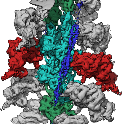 emd_8165.png
 http://ftp.pdbj.org/pub/emdb/structures/EMD-8165


 Z (Sec.)
 Y (Row.)
 X (Col.)


 試料の構成要素
 Spodoptera frugiperda (ツマジロクサヨトウ)
 解析
解析 電子顕微鏡法
 FIELD EMISSION GUN