 Movie
Movie Controller


+ Open data

- Basic information
| Entry | Database: PDB / ID: 4pb6 | ||||||
|---|---|---|---|---|---|---|---|
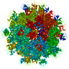
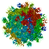
| Title | Feline calicivirus VP1 T=1 virus-like particle | ||||||
 Components Components | VP1 | ||||||
 Keywords Keywords | VIRUS / viral capsid protein / virus-like particle | ||||||
| Function / homology | T=3 icosahedral viral capsid / Calicivirus coat protein / Calicivirus coat protein / Picornavirus/Calicivirus coat protein / Viral coat protein subunit / host cell cytoplasm / Capsid protein VP1 Function and homology information Function and homology information | ||||||
| Biological species |  Feline calicivirus Feline calicivirus | ||||||
| Method |  X-RAY DIFFRACTION / SYNCHROTRON / MOLECULAR REPLACEMENT / Resolution: 8 Å X-RAY DIFFRACTION / SYNCHROTRON / MOLECULAR REPLACEMENT / Resolution: 8 Å | ||||||
 Authors Authors | Burmeister, W.P. / Buisson, M. | ||||||
 Citation Citation | Journal: PLoS One / Year: 2015 Title: Structure determination of feline calicivirus virus-like particles in the context of a pseudo-octahedral arrangement. Authors: Wim P Burmeister / Marlyse Buisson / Leandro F Estrozi / Guy Schoehn / Olivier Billet / Zahia Hannas / Cécile Sigoillot / Hervé Poulet /  Abstract: The vesivirus feline calicivirus (FCV) is a positive strand RNA virus encapsidated by an icosahedral T=3 shell formed by the viral VP1 protein. Upon its expression in the insect cell - baculovirus ...The vesivirus feline calicivirus (FCV) is a positive strand RNA virus encapsidated by an icosahedral T=3 shell formed by the viral VP1 protein. Upon its expression in the insect cell - baculovirus system in the context of vaccine development, two types of virus-like particles (VLPs) were formed, a majority built of 60 subunits (T=1) and a minority probably built of 180 subunits (T=3). The structure of the small particles was determined by x-ray crystallography at 0.8 nm resolution helped by cryo-electron microscopy in order to understand their formation. Cubic crystals belonged to space group P213. Their self-rotation function showed the presence of an octahedral pseudo-symmetry similar to the one described previously by Agerbandje and co-workers for human parvovirus VLPs. The crystal structure could be solved starting from the published VP1 structure in the context of the T=3 viral capsid. In contrast to viral capsids, where the capsomers are interlocked by the exchange of the N-terminal arm (NTA) domain, this domain is disordered in the T=1 capsid of the VLPs. Furthermore it is prone to proteolytic cleavage. The relative orientation of P (protrusion) and S (shell) domains is alerted so as to fit VP1 to the smaller T=1 particle whereas the intermolecular contacts around 2-fold, 3-fold and 5-fold axes are conserved. By consequence the surface of the VLP is very similar compared to the viral capsid and suggests a similar antigenicity. The knowledge of the structure of the VLPs will help to improve their stability, in respect to a use for vaccination. | ||||||
| History |
|
- Structure visualization
| Structure viewer | Molecule: MolmilJmol/JSmol |
|---|
- Downloads & links
-Download
| PDBx/mmCIF format | 4pb6.cif.gz | 1.6 MB | Display | PDBx/mmCIF format |
|---|---|---|---|---|
| PDB format | pdb4pb6.ent.gz | 1.3 MB | Display | PDB format |
| PDBx/mmJSON format | 4pb6.json.gz | Tree view | PDBx/mmJSON format | |
| Others |  Other downloads Other downloads |
-Validation report
| Arichive directory | https://data.pdbj.org/pub/pdb/validation_reports/pb/4pb6ftp://data.pdbj.org/pub/pdb/validation_reports/pb/4pb6 | HTTPS FTP |
|---|
-Related structure data
| Related structure data |  2823C  3m8lS S: Starting model for refinement C: citing same article ( |
|---|---|
| Similar structure data |


-Links
 PDBj
PDBj

- Assembly
| Deposited unit | 
| ||||||||||||
|---|---|---|---|---|---|---|---|---|---|---|---|---|---|
| 1 | 
| ||||||||||||
| 2 |
| ||||||||||||
| 3 | 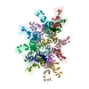
| ||||||||||||
| Unit cell |
| ||||||||||||
| Symmetry | Point symmetry: (Schoenflies symbol: C3 (3 fold cyclic)) | ||||||||||||
| Noncrystallographic symmetry (NCS) | NCS oper:
|
-Components
| #1: Protein | Mass: 59297.613 Da / Num. of mol.: 20 Source method: isolated from a genetically manipulated source Source: (gene. exp.) Feline calicivirus / Strain: Merial100869 / Gene: VP1 / Cell line (production host): Sf9 / Production host:   Spodoptera frugiperda (fall armyworm) / References: UniProt: A0A0J9X1Z3*PLUS Spodoptera frugiperda (fall armyworm) / References: UniProt: A0A0J9X1Z3*PLUS |
|---|
-Experimental details
-Experiment
| Experiment | Method: X-RAY DIFFRACTION |
|---|
- Sample preparation
| Crystal | Density Matthews: 3.24 Å3/Da / Density % sol: 62 % / Description: tetrahedra or octahedra |
|---|---|
| Crystal grow | Temperature: 293 K / Method: vapor diffusion, hanging drop / pH: 7 Details: reservoir solution: 7 - 10 % PEG 6000, 0.1 M Hepes pH 7, 0.5 - 1 M LiCl protein in: 20 mM MES pH 6, 200 mM NaCl at 3.4 mg/ml |
-Data collection
| Diffraction | Mean temperature: 100 K |
|---|---|
| Diffraction source | Source: SYNCHROTRON / Site: ESRF / Beamline: ID23-2 / Wavelength: 0.9185 Å |
| Detector | Type: ADSC QUANTUM 4 / Detector: CCD / Date: Feb 15, 2010 |
| Radiation | Protocol: SINGLE WAVELENGTH / Monochromatic (M) / Laue (L): M / Scattering type: x-ray |
| Radiation wavelength | Wavelength: 0.9185 Å / Relative weight: 1 |
| Reflection | Resolution: 8→67.2 Å / Num. all: 14809 / Num. obs: 14809 / % possible obs: 87.22 % / Redundancy: 5.5 % / Rmerge(I) obs: 0.24 / Rsym value: 0.24 / Net I/av σ(I): 6.64 / Net I/σ(I): 3.07 |
| Reflection shell | Resolution: 8→8.43 Å / Redundancy: 1.82 % / Rmerge(I) obs: 1.1 / Mean I/σ(I) obs: 0.69 / % possible all: 61.5 |
- Processing
| Software |
| |||||||||||||||||||||
|---|---|---|---|---|---|---|---|---|---|---|---|---|---|---|---|---|---|---|---|---|---|---|
| Refinement | Method to determine structure: MOLECULAR REPLACEMENT Starting model: pdb 3m8l and model builting Resolution: 8→65.51 Å / Cor.coef. Fo:Fc: 0.503 / Cor.coef. Fo:Fc free: 0.42 / SU B: 0.003 / SU ML: 0 / Cross valid method: THROUGHOUT / ESU R: 8.911 / ESU R Free: 9.346 / Stereochemistry target values: MAXIMUM LIKELIHOOD / Details: HYDROGENS HAVE BEEN USED IF PRESENT IN THE INPUT
| |||||||||||||||||||||
| Solvent computation | Ion probe radii: 0.8 Å / Shrinkage radii: 0.8 Å / VDW probe radii: 1.2 Å / Solvent model: BABINET MODEL WITH MASK | |||||||||||||||||||||
| Displacement parameters | Biso mean: 94.995 Å2
| |||||||||||||||||||||
| Refinement step | Cycle: 1 / Resolution: 8→65.51 Å
| |||||||||||||||||||||
| LS refinement shell | Refine-ID: X-RAY DIFFRACTION
|
