 Movie
Movie Controller

[English] 日本語
 Yorodumi
- EMDB-8047: Electron tomographic structure of an individual human plasma VLDL... -
+ Open data

- Basic information
| Entry | Database: EMDB / ID: EMD-8047 | |||||||||
|---|---|---|---|---|---|---|---|---|---|---|
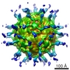
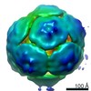
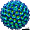
| Title | Electron tomographic structure of an individual human plasma VLDL particle (No.006) | |||||||||
 Map data Map data | human plasma VLDL particle | |||||||||
 Sample Sample |
| |||||||||
| Biological species |  Homo sapiens (human) Homo sapiens (human) | |||||||||
| Method | electron tomography / cryo EM / Resolution: 50.0 Å | |||||||||
 Authors Authors | Yu Y / Kuang Y / Lei D / Zhai X / Krauss R / Ren G | |||||||||
| Funding support |  United States, 2 items United States, 2 items
| |||||||||
 Citation Citation | Journal: J Lipid Res / Year: 2016 Title: Polyhedral 3D structure of human plasma very low density lipoproteins by individual particle cryo-electron tomography1. Authors: Yadong Yu / Yu-Lin Kuang / Dongsheng Lei / Xiaobo Zhai / Meng Zhang / Ronald M Krauss / Gang Ren / Abstract: Human VLDLs assembled in the liver and secreted into the circulation supply energy to peripheral tissues. VLDL lipolysis yields atherogenic LDLs and VLDL remnants that strongly correlate with CVD. ...Human VLDLs assembled in the liver and secreted into the circulation supply energy to peripheral tissues. VLDL lipolysis yields atherogenic LDLs and VLDL remnants that strongly correlate with CVD. Although the composition of VLDL particles has been well-characterized, their 3D structure is elusive because of their variations in size, heterogeneity in composition, structural flexibility, and mobility in solution. Here, we employed cryo-electron microscopy and individual-particle electron tomography to study the 3D structure of individual VLDL particles (without averaging) at both below and above their lipid phase transition temperatures. The 3D reconstructions of VLDL and VLDL bound to antibodies revealed an unexpected polyhedral shape, in contrast to the generally accepted model of a spherical emulsion-like particle. The smaller curvature of surface lipids compared with HDL may also reduce surface hydrophobicity, resulting in lower binding affinity to the hydrophobic distal end of the N-terminal β-barrel domain of cholesteryl ester transfer protein (CETP) compared with HDL. The directional binding of CETP to HDL and VLDL may explain the function of CETP in transferring TGs and cholesteryl esters between these particles. This first visualization of the 3D structure of VLDL could improve our understanding of the role of VLDL in atherogenesis. | |||||||||
| History |
|
- Structure visualization
| Movie |
 Movie viewer Movie viewer |
|---|---|
| Structure viewer | EM map: SurfViewMolmilJmol/JSmol |
| Supplemental images |
 UCSF Chimera
- Downloads & links
-EMDB archive
| Map data | emd_8047.map.gz | 48.5 MB | EMDB map data format | |
|---|---|---|---|---|
| Header (meta data) | emd-8047-v30.xmlemd-8047.xml | 15.8 KB 15.8 KB | Display Display | EMDB header |
| Images | 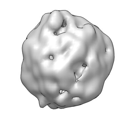 emd_8047.png emd_8047.png | 57.7 KB | ||
| Archive directory |  http://ftp.pdbj.org/pub/emdb/structures/EMD-8047ftp://ftp.pdbj.org/pub/emdb/structures/EMD-8047 http://ftp.pdbj.org/pub/emdb/structures/EMD-8047ftp://ftp.pdbj.org/pub/emdb/structures/EMD-8047 | HTTPS FTP |
-Related structure data
| Related structure data | 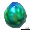 8042C 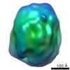 8043C 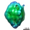 8044C 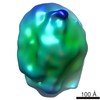 8045C 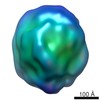 8046C 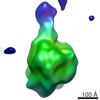 8048C 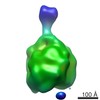 8049C 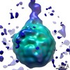 8050C 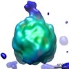 8051C 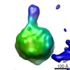 8052C 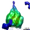 8053C C: citing same article ( |
|---|---|
| Similar structure data |

-Links
| EMDB pages | EMDB (EBI/PDBe) / EMDataResource |
|---|
-Map
| File | Download / File: emd_8047.map.gz / Format: CCP4 / Size: 52.7 MB / Type: IMAGE STORED AS FLOATING POINT NUMBER (4 BYTES) | ||||||||||||||||||||||||||||||||||||||||||||||||||||||||||||||||||||
|---|---|---|---|---|---|---|---|---|---|---|---|---|---|---|---|---|---|---|---|---|---|---|---|---|---|---|---|---|---|---|---|---|---|---|---|---|---|---|---|---|---|---|---|---|---|---|---|---|---|---|---|---|---|---|---|---|---|---|---|---|---|---|---|---|---|---|---|---|---|
| Annotation | human plasma VLDL particle | ||||||||||||||||||||||||||||||||||||||||||||||||||||||||||||||||||||
| Projections & slices | Image control
Images are generated by Spider. | ||||||||||||||||||||||||||||||||||||||||||||||||||||||||||||||||||||
| Voxel size | X=Y=Z: 2.4 Å | ||||||||||||||||||||||||||||||||||||||||||||||||||||||||||||||||||||
| Density |
| ||||||||||||||||||||||||||||||||||||||||||||||||||||||||||||||||||||
| Symmetry | Space group: 1 | ||||||||||||||||||||||||||||||||||||||||||||||||||||||||||||||||||||
| Details | EMDB XML:
CCP4 map header:
| ||||||||||||||||||||||||||||||||||||||||||||||||||||||||||||||||||||
 Z (Sec.)
 Y (Row.)
 X (Col.)


-Supplemental data
- Sample components
-Entire : Human Plasma Very-Low-Density Lipoprotein
| Entire | Name: Human Plasma Very-Low-Density Lipoprotein |
|---|---|
| Components |
|
-Supramolecule #1: Human Plasma Very-Low-Density Lipoprotein
| Supramolecule | Name: Human Plasma Very-Low-Density Lipoprotein / type: complex / ID: 1 / Parent: 0 Details: VLDL isolated from human plasma by density gradient centrifugation. |
|---|---|
| Source (natural) | Organism: Homo sapiens (human) / Tissue: blood |
| Molecular weight | Experimental: 14.0 MDa |
-Experimental details
-Structure determination
| Method | cryo EM |
|---|---|
 Processing Processing | electron tomography |
| Aggregation state | particle |
-Sample preparation
| Concentration | 0.4 mg/mL | |||||||||||||||
|---|---|---|---|---|---|---|---|---|---|---|---|---|---|---|---|---|
| Buffer | pH: 7.3 Component:
Details: DPBS | |||||||||||||||
| Grid | Model: EMS Lacey carbon / Material: COPPER / Mesh: 200 / Support film - Material: CARBON / Support film - topology: LACEY / Support film - Film thickness: 50.0 nm / Pretreatment - Type: GLOW DISCHARGE / Pretreatment - Atmosphere: AIR / Pretreatment - Pressure: 0.039 kPa | |||||||||||||||
| Vitrification | Cryogen name: ETHANE / Chamber humidity: 90 % / Chamber temperature: 277 K / Instrument: LEICA EM GP Details: 3 microliter of specimen blotted for 3 seconds before plunging. | |||||||||||||||
| Details | This sample was monodisperse. | |||||||||||||||
| Sectioning | Other: NO SECTIONING |
- Electron microscopy
| Microscope | ZEISS LIBRA120PLUS |
|---|---|
| Temperature | Min: 90.0 K / Max: 93.0 K |
| Specialist optics | Energy filter - Name: Zeiss in-column Omega / Energy filter - Lower energy threshold: 0 eV / Energy filter - Upper energy threshold: 20 eV |
| Image recording | Film or detector model: GATAN ULTRASCAN 4000 (4k x 4k) / Digitization - Dimensions - Width: 4096 pixel / Digitization - Dimensions - Height: 4096 pixel / Digitization - Sampling interval: 15.0 µm / Number grids imaged: 1 / Number real images: 85 / Average exposure time: 0.5 sec. / Average electron dose: 1.8 e/Å2 Details: Images were collected by using the Gatan Tomography module of Digital Micrograph. XY tracking and focusing were done manually. |
| Electron beam | Acceleration voltage: 120 kV / Electron source: LAB6 |
| Electron optics | C2 aperture diameter: 75.0 µm / Calibrated defocus max: 2.5 µm / Calibrated defocus min: 1.5 µm / Calibrated magnification: 50000 / Illumination mode: FLOOD BEAM / Imaging mode: BRIGHT FIELD / Cs: 2.2 mm / Nominal defocus max: 2.5 µm / Nominal defocus min: 1.5 µm / Nominal magnification: 50000 |
| Sample stage | Specimen holder model: GATAN 626 SINGLE TILT LIQUID NITROGEN CRYO TRANSFER HOLDER Cooling holder cryogen: NITROGEN |
-Image processing
| Details | Defects in images such as bad pixels and X-rays were removed prior to alignment and 3D reconstruction. |
|---|---|
| Final reconstruction | Algorithm: BACK PROJECTION / Resolution.type: BY AUTHOR / Resolution: 50.0 Å / Resolution method: FSC 0.5 CUT-OFF / Software - Name: IPET Details: The 3D reconstruction was conducted by using Individual Particle Electron Tomography (IPET) Number images used: 85 |
| CTF correction | Software - Name: TOMOCTF (ver. V. October 2012) |
