 Movie
Movie Controller


+ Open data

- Basic information
| Entry | Database: EMDB / ID: EMD-1942 | |||||||||
|---|---|---|---|---|---|---|---|---|---|---|
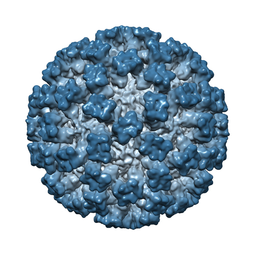
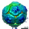
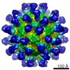
| Title | Feline Calicivirus strain F9 | |||||||||
 Map data Map data | Three-dimensional reconstruction of Feline Calicivirus strain f9 at 9 Angstroms resolution | |||||||||
 Sample Sample |
| |||||||||
 Keywords Keywords | feline / calicivirus / virus / virion | |||||||||
| Biological species |  Feline calicivirus Feline calicivirus | |||||||||
| Method | single particle reconstruction / cryo EM / Resolution: 8.9 Å | |||||||||
 Authors Authors | Bhella D / Goodfellow IG | |||||||||
 Citation Citation | Journal: J Virol / Year: 2011 Title: The cryo-electron microscopy structure of feline calicivirus bound to junctional adhesion molecule A at 9-angstrom resolution reveals receptor-induced flexibility and two distinct ...Title: The cryo-electron microscopy structure of feline calicivirus bound to junctional adhesion molecule A at 9-angstrom resolution reveals receptor-induced flexibility and two distinct conformational changes in the capsid protein VP1. Authors: David Bhella / Ian G Goodfellow /  Abstract: Caliciviridae are small icosahedral positive-sense RNA-containing viruses and include the human noroviruses, a leading cause of infectious acute gastroenteritis and feline calicivirus (FCV), which ...Caliciviridae are small icosahedral positive-sense RNA-containing viruses and include the human noroviruses, a leading cause of infectious acute gastroenteritis and feline calicivirus (FCV), which causes respiratory illness and stomatitis in cats. FCV attachment and entry is mediated by feline junctional adhesion molecule A (fJAM-A), which binds to the outer face of the capsomere, inducing a conformational change in the capsid that may be important for viral uncoating. Here we present the results of our structural investigation of the virus-receptor interaction and ensuing conformational changes. Cryo-electron microscopy and three-dimensional image reconstruction were used to solve the structure of the virus decorated with a soluble fragment of the receptor at subnanometer resolution. In initial reconstructions, the P domains of the capsid protein VP1 and fJAM-A were poorly resolved. Sorting experiments led to improved reconstructions of the FCV-fJAM-A complex both before and after the induced conformational change, as well as in three transition states. These data showed that the P domain becomes flexible following fJAM-A binding, leading to a loss of icosahedral symmetry. Furthermore, two distinct conformational changes were seen; an anticlockwise rotation of up to 15° of the P domain was observed in the AB dimers, while tilting of the P domain away from the icosahedral 2-fold axis was seen in the CC dimers. A list of putative contact residues was calculated by fitting high-resolution coordinates for fJAM-A and VP1 to the reconstructed density maps, highlighting regions in both virus and receptor important for virus attachment and entry. | |||||||||
| History |
|
- Structure visualization
| Movie |
 Movie viewer Movie viewer |
|---|---|
| Structure viewer | EM map: SurfViewMolmilJmol/JSmol |
| Supplemental images |
- Downloads & links
-EMDB archive
| Map data | emd_1942.map.gz | 29.5 MB | EMDB map data format | |
|---|---|---|---|---|
| Header (meta data) | emd-1942-v30.xmlemd-1942.xml | 8.8 KB 8.8 KB | Display Display | EMDB header |
| Images |  EMD-1942.jpg EMD-1942.jpg | 184.9 KB | ||
| Archive directory |  http://ftp.pdbj.org/pub/emdb/structures/EMD-1942ftp://ftp.pdbj.org/pub/emdb/structures/EMD-1942 http://ftp.pdbj.org/pub/emdb/structures/EMD-1942ftp://ftp.pdbj.org/pub/emdb/structures/EMD-1942 | HTTPS FTP |
-Related structure data
| Related structure data |  1943C  1944C  1945C  1946C  1947C  1948C C: citing same article ( |
|---|---|
| Similar structure data |

-Links
| EMDB pages | EMDB (EBI/PDBe) / EMDataResource |
|---|
-Map
| File | Download / File: emd_1942.map.gz / Format: CCP4 / Size: 61.8 MB / Type: IMAGE STORED AS FLOATING POINT NUMBER (4 BYTES) | ||||||||||||||||||||||||||||||||||||||||||||||||||||||||||||||||||||
|---|---|---|---|---|---|---|---|---|---|---|---|---|---|---|---|---|---|---|---|---|---|---|---|---|---|---|---|---|---|---|---|---|---|---|---|---|---|---|---|---|---|---|---|---|---|---|---|---|---|---|---|---|---|---|---|---|---|---|---|---|---|---|---|---|---|---|---|---|---|
| Annotation | Three-dimensional reconstruction of Feline Calicivirus strain f9 at 9 Angstroms resolution | ||||||||||||||||||||||||||||||||||||||||||||||||||||||||||||||||||||
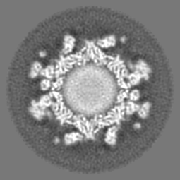
| Projections & slices | Image control
Images are generated by Spider. | ||||||||||||||||||||||||||||||||||||||||||||||||||||||||||||||||||||
| Voxel size | X=Y=Z: 2.06 Å | ||||||||||||||||||||||||||||||||||||||||||||||||||||||||||||||||||||
| Density |
| ||||||||||||||||||||||||||||||||||||||||||||||||||||||||||||||||||||
| Symmetry | Space group: 1 | ||||||||||||||||||||||||||||||||||||||||||||||||||||||||||||||||||||
| Details | EMDB XML:
CCP4 map header:
| ||||||||||||||||||||||||||||||||||||||||||||||||||||||||||||||||||||
 Z (Sec.)
 Y (Row.)
 X (Col.)


-Supplemental data
- Sample components
-Entire : Feline Calicivirus
| Entire | Name: Feline Calicivirus |
|---|---|
| Components |
|
-Supramolecule #1000: Feline Calicivirus
| Supramolecule | Name: Feline Calicivirus / type: sample / ID: 1000 / Oligomeric state: T3 icosahedral capsid / Number unique components: 1 |
|---|---|
| Molecular weight | Theoretical: 10.7 MDa |
-Supramolecule #1: Feline calicivirus
| Supramolecule | Name: Feline calicivirus / type: virus / ID: 1 / Name.synonym: Feline calicivirus / NCBI-ID: 11978 / Sci species name: Feline calicivirus / Virus type: VIRION / Virus isolate: STRAIN / Virus enveloped: No / Virus empty: No / Syn species name: Feline calicivirus |
|---|---|
| Host (natural) | Organism:  |
| Molecular weight | Theoretical: 10.7 MDa |
| Virus shell | Shell ID: 1 / Name: VP1 / Diameter: 415 Å / T number (triangulation number): 3 |
-Experimental details
-Structure determination
| Method | cryo EM |
|---|---|
 Processing Processing | single particle reconstruction |
| Aggregation state | particle |
-Sample preparation
| Buffer | pH: 7.4 / Details: PBS |
|---|---|
| Grid | Details: 400 mesh R2/2 C-flat |
| Vitrification | Cryogen name: ETHANE / Chamber humidity: 100 % / Instrument: OTHER / Details: Vitrification instrument: Vitrobot / Method: blot for five seconds before plunging |
- Electron microscopy
| Microscope | JEOL 2200FS |
|---|---|
| Temperature | Average: 97 K |
| Alignment procedure | Legacy - Astigmatism: corrected at 100k times mag in digital micrograph |
| Specialist optics | Energy filter - Name: JEOL / Energy filter - Lower energy threshold: 0.0 eV / Energy filter - Upper energy threshold: 20.0 eV |
| Image recording | Category: CCD / Film or detector model: GATAN ULTRASCAN 4000 (4k x 4k) / Average electron dose: 10 e/Å2 |
| Electron beam | Acceleration voltage: 200 kV / Electron source:  FIELD EMISSION GUN FIELD EMISSION GUN |
| Electron optics | Illumination mode: FLOOD BEAM / Imaging mode: BRIGHT FIELD / Cs: 2.0 mm / Nominal defocus max: 3.5 µm / Nominal defocus min: 0.8 µm / Nominal magnification: 100000 |
| Sample stage | Specimen holder: Side entry / Specimen holder model: GATAN LIQUID NITROGEN |
-Image processing
| CTF correction | Details: each particle corrected using bsoft |
|---|---|
| Final reconstruction | Applied symmetry - Point group: I (icosahedral) / Algorithm: OTHER / Resolution.type: BY AUTHOR / Resolution: 8.9 Å / Resolution method: FSC 0.5 CUT-OFF / Software - Name: em3dr2 / Number images used: 6802 |
