 Movie
Movie Controller

[English] 日本語
 Yorodumi
- EMDB-7979: Cytoplasmic domain of human type 3 1,4,5-inositol trisphosphate r... -
+ Open data

- Basic information
| Entry | Database: EMDB / ID: EMD-7979 | |||||||||
|---|---|---|---|---|---|---|---|---|---|---|
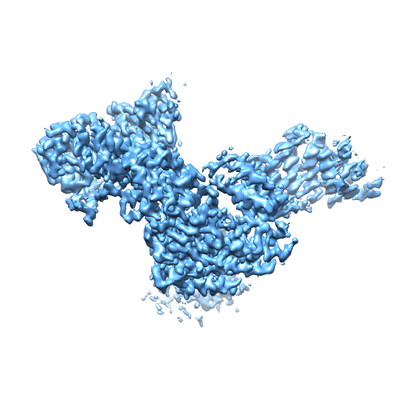
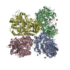
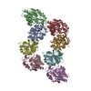
| Title | Cytoplasmic domain of human type 3 1,4,5-inositol trisphosphate receptor in a ligand-free state | |||||||||
 Map data Map data | Cytoplasmic domain focused refinement of human type 3 1,4,5-inositol trisphosphate receptor in a ligand-free state | |||||||||
 Sample Sample |
| |||||||||
| Biological species |  Homo sapiens (human) Homo sapiens (human) | |||||||||
| Method | single particle reconstruction / cryo EM / Resolution: 3.69 Å | |||||||||
 Authors Authors | Hite RK / Paknejad N | |||||||||
 Citation Citation | Journal: Nat Struct Mol Biol / Year: 2018 Title: Structural basis for the regulation of inositol trisphosphate receptors by Ca and IP. Authors: Navid Paknejad / Richard K Hite /  Abstract: Inositol trisphosphate receptors (IPRs) are ubiquitous Ca-permeable channels that mediate release of Ca from the endoplasmic reticulum, thereby regulating numerous processes including cell division, ...Inositol trisphosphate receptors (IPRs) are ubiquitous Ca-permeable channels that mediate release of Ca from the endoplasmic reticulum, thereby regulating numerous processes including cell division, cell death, differentiation and fertilization. IPRs are jointly activated by inositol trisphosphate (IP) and their permeant ion, Ca. At high concentrations, however, Ca inhibits activity, ensuring precise spatiotemporal control over intracellular Ca. Despite extensive characterization of IPR, the mechanisms through which these molecules control channel gating have remained elusive. Here, we present structures of full-length human type 3 IPRs in ligand-bound and ligand-free states. Multiple IP-bound structures demonstrate that the large cytoplasmic domain provides a platform for propagation of long-range conformational changes to the ion-conduction gate. Structures in the presence of Ca reveal two Ca-binding sites that induce the disruption of numerous interactions between subunits, thereby inhibiting IPR. These structures thus provide a mechanistic basis for beginning to understand the regulation of IPR. | |||||||||
| History |
|
- Structure visualization
| Movie |
 Movie viewer Movie viewer |
|---|---|
| Structure viewer | EM map: SurfViewMolmilJmol/JSmol |
| Supplemental images |
- Downloads & links
-EMDB archive
| Map data | emd_7979.map.gz | 190.2 MB | EMDB map data format | |
|---|---|---|---|---|
| Header (meta data) | emd-7979-v30.xmlemd-7979.xml | 13.7 KB 13.7 KB | Display Display | EMDB header |
| Images |  emd_7979.png emd_7979.png | 103.1 KB | ||
| Archive directory |  http://ftp.pdbj.org/pub/emdb/structures/EMD-7979ftp://ftp.pdbj.org/pub/emdb/structures/EMD-7979 http://ftp.pdbj.org/pub/emdb/structures/EMD-7979ftp://ftp.pdbj.org/pub/emdb/structures/EMD-7979 | HTTPS FTP |
-Validation report
| Summary document | emd_7979_validation.pdf.gz | 78.2 KB | Display | EMDB validaton report |
|---|---|---|---|---|
| Full document | emd_7979_full_validation.pdf.gz | 77.3 KB | Display | |
| Data in XML | emd_7979_validation.xml.gz | 494 B | Display | |
| Arichive directory | https://ftp.pdbj.org/pub/emdb/validation_reports/EMD-7979ftp://ftp.pdbj.org/pub/emdb/validation_reports/EMD-7979 | HTTPS FTP |
-Related structure data
| Related structure data |  7978C  7980C  7981C  7982C 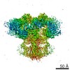 7983C 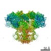 7984C 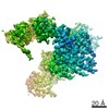 7985C 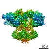 7986C 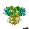 7987C 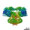 7988C 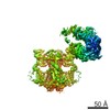 7989C 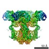 7990C 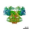 7991C 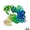 7992C 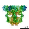 7993C 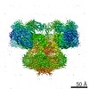 7994C 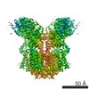 7995C 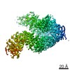 7996C 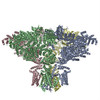 6dqjC 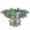 6dqnC 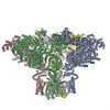 6dqsC 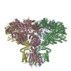 6dqvC 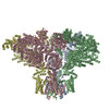 6dqzC 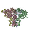 6dr0C  6dr2C  6draC  6drcC C: citing same article ( |
|---|---|
| Similar structure data |


-Links
| EMDB pages | EMDB (EBI/PDBe) / EMDataResource |
|---|
-Map
| File | Download / File: emd_7979.map.gz / Format: CCP4 / Size: 216 MB / Type: IMAGE STORED AS FLOATING POINT NUMBER (4 BYTES) | ||||||||||||||||||||||||||||||||||||||||||||||||||||||||||||||||||||
|---|---|---|---|---|---|---|---|---|---|---|---|---|---|---|---|---|---|---|---|---|---|---|---|---|---|---|---|---|---|---|---|---|---|---|---|---|---|---|---|---|---|---|---|---|---|---|---|---|---|---|---|---|---|---|---|---|---|---|---|---|---|---|---|---|---|---|---|---|---|
| Annotation | Cytoplasmic domain focused refinement of human type 3 1,4,5-inositol trisphosphate receptor in a ligand-free state | ||||||||||||||||||||||||||||||||||||||||||||||||||||||||||||||||||||
| Projections & slices | Image control
Images are generated by Spider. | ||||||||||||||||||||||||||||||||||||||||||||||||||||||||||||||||||||
| Voxel size | X=Y=Z: 1.088 Å | ||||||||||||||||||||||||||||||||||||||||||||||||||||||||||||||||||||
| Density |
| ||||||||||||||||||||||||||||||||||||||||||||||||||||||||||||||||||||
| Symmetry | Space group: 1 | ||||||||||||||||||||||||||||||||||||||||||||||||||||||||||||||||||||
| Details | EMDB XML:
CCP4 map header:
| ||||||||||||||||||||||||||||||||||||||||||||||||||||||||||||||||||||
 Z (Sec.)
 Y (Row.)
 X (Col.)


-Supplemental data
- Sample components
-Entire : human type 3 inositol 1,4,5-trisphosphate receptor
| Entire | Name: human type 3 inositol 1,4,5-trisphosphate receptor |
|---|---|
| Components |
|
-Supramolecule #1: human type 3 inositol 1,4,5-trisphosphate receptor
| Supramolecule | Name: human type 3 inositol 1,4,5-trisphosphate receptor / type: organelle_or_cellular_component / ID: 1 / Parent: 0 / Macromolecule list: #1 |
|---|---|
| Source (natural) | Organism: Homo sapiens (human) |
| Recombinant expression | Organism: Homo sapiens (human) / Recombinant strain: HEK 293S GnTi / Recombinant plasmid: BacMam |
-Experimental details
-Structure determination
| Method | cryo EM |
|---|---|
 Processing Processing | single particle reconstruction |
| Aggregation state | particle |
-Sample preparation
| Concentration | 8 mg/mL | ||||||||||||||||||
|---|---|---|---|---|---|---|---|---|---|---|---|---|---|---|---|---|---|---|---|
| Buffer | pH: 8 Component:
Details: 150mM Nacl 50mM Tris-HCl, pH 8.0 2mM DTT 0.06% Digitonin 5mM EGTA | ||||||||||||||||||
| Grid | Model: Quantifoil R1.2/1.3 / Material: GOLD / Mesh: 400 / Support film - Material: CARBON / Support film - topology: HOLEY ARRAY | ||||||||||||||||||
| Vitrification | Cryogen name: ETHANE / Chamber humidity: 100 % / Chamber temperature: 293 K / Instrument: FEI VITROBOT MARK IV / Details: Blot for 2 seconds prior to freezing. | ||||||||||||||||||
| Details | ligand-free human type 3 inositol 1,4,5-trisphosphate receptor in detergent micelles |
- Electron microscopy
| Microscope | FEI TITAN KRIOS |
|---|---|
| Specialist optics | Spherical aberration corrector: FEI Cs corrector / Energy filter - Name: GIF Bioquantum / Energy filter - Slit width: 20 eV |
| Image recording | Film or detector model: GATAN K2 SUMMIT (4k x 4k) / Detector mode: SUPER-RESOLUTION / Digitization - Dimensions - Width: 7420 pixel / Digitization - Dimensions - Height: 7676 pixel / Digitization - Sampling interval: 5.0 µm / Digitization - Frames/image: 1-40 / Number grids imaged: 1 / Number real images: 1801 / Average exposure time: 8.0 sec. / Average electron dose: 60.0 e/Å2 |
| Electron beam | Acceleration voltage: 300 kV / Electron source:  FIELD EMISSION GUN FIELD EMISSION GUN |
| Electron optics | C2 aperture diameter: 70.0 µm / Illumination mode: FLOOD BEAM / Imaging mode: BRIGHT FIELD / Cs: 0.0 mm / Nominal defocus min: 1.0 µm / Nominal magnification: 105000 |
| Sample stage | Specimen holder model: FEI TITAN KRIOS AUTOGRID HOLDER / Cooling holder cryogen: NITROGEN |
| Experimental equipment |  Model: Titan Krios / Image courtesy: FEI Company |
