National Institutes of Health/National Institute of General Medical Sciences
R01 GM102474
米国
National Institutes of Health/National Institute of General Medical Sciences
U24GM116787
米国
引用
ジャーナル: Sci Rep / 年: 2020 タイトル: Complexity and ultrastructure of infectious extracellular vesicles from cells infected by non-enveloped virus. 著者: Jie E Yang / Evan D Rossignol / Deborah Chang / Joseph Zaia / Isaac Forrester / Kiran Raja / Holly Winbigler / Daniela Nicastro / William T Jackson / Esther Bullitt / 要旨: Enteroviruses support cell-to-cell viral transmission prior to their canonical lytic spread of virus. Poliovirus (PV), a prototype for human pathogenic positive-sense RNA enteroviruses, and ...Enteroviruses support cell-to-cell viral transmission prior to their canonical lytic spread of virus. Poliovirus (PV), a prototype for human pathogenic positive-sense RNA enteroviruses, and picornaviruses in general, transport multiple virions en bloc via infectious extracellular vesicles, 100~1000 nm in diameter, secreted from host cells. Using biochemical and biophysical methods we identify multiple components in secreted microvesicles, including mature PV virions; positive-sense genomic and negative-sense replicative, template viral RNA; essential viral replication proteins; and cellular proteins. Using cryo-electron tomography, we visualize the near-native three-dimensional architecture of secreted infectious microvesicles containing both virions and a unique morphological component that we describe as a mat-like structure. While the composition of these mat-like structures is not yet known, based on our biochemical data they are expected to be comprised of unencapsidated RNA and proteins. In addition to infectious microvesicles, CD9-positive exosomes released from PV-infected cells are also infectious and transport virions. Thus, our data show that, prior to cell lysis, non-enveloped viruses are secreted within infectious vesicles that also transport viral unencapsidated RNAs, viral and host proteins. Understanding the structure and function of these infectious particles helps elucidate the mechanism by which extracellular vesicles contribute to the spread of non-enveloped virus infection.
#20 - 2001年8月 ポリオウイルスとライノウイルス (Poliovirus and Rhinovirus) 類似性 (1)
-
マップ
ファイル
ダウンロード / ファイル: emd_7881.map.gz / 形式: CCP4 / 大きさ: 937.4 MB / タイプ: IMAGE STORED AS SIGNED BYTE
注釈
Subcellular fractionation of viral replication membranes from poliovirus-infected HeLa cells at 5 hpi
ボクセルのサイズ
X=Y=Z: 8.2 Å
密度
最小 - 最大
-128 - 127
平均 (標準偏差)
31.97968 (±117.889206)
対称性
空間群: 1
詳細
EMDB XML:
マップ形状
Axis order
X
Y
Z
Origin
0
0
208
サイズ
1602
1404
437
Spacing
1404
1602
437
セル
A: 11512.8 Å / B: 13136.399 Å / C: 3583.4 Å α=β=γ: 90.0 °
CCP4マップ ヘッダ情報:
mode
envelope stored as signed bytes (from -128 lowest to 127 highest)
Å/pix. X/Y/Z
8.2
8.1999993757803
8.2
M x/y/z
1404
1602
437
origin x/y/z
0.000
0.000
0.000
length x/y/z
11512.800
13136.399
3583.400
α/β/γ
90.000
90.000
90.000
start NX/NY/NZ
0
0
0
NX/NY/NZ
280
280
280
MAP C/R/S
1
2
3
start NC/NR/NS
0
0
208
NC/NR/NS
1404
1602
437
D min/max/mean
-128.000
127.000
31.980
-
添付データ
-
試料の構成要素
-
全体 : Human poliovirus 1 Mahoney
全体
名称: Human poliovirus 1 Mahoney (ポリオウイルス)
要素
ウイルス: Human poliovirus 1 Mahoney (ポリオウイルス)
-
超分子 #1: Human poliovirus 1 Mahoney
超分子
名称: Human poliovirus 1 Mahoney / タイプ: virus / ID: 1 / 親要素: 0 詳細: virion-containing microvesicles collected from supernatant of poliovirus-infected cells at 8 hours post-infection NCBI-ID: 12081 / 生物種: Human poliovirus 1 Mahoney / ウイルスタイプ: VIRUS-LIKE PARTICLE / ウイルス・単離状態: STRAIN / ウイルス・エンベロープ: No / ウイルス・中空状態: No
宿主
生物種: Homo sapiens (ヒト)
-
実験情報
-
構造解析
手法
クライオ電子顕微鏡法
解析
電子線トモグラフィー法
試料の集合状態
particle
-
試料調製
濃度
1.5 mg/mL
緩衝液
pH: 7 / 詳細: 1% glutaraldehyde in 1x PBS
グリッド
材質: COPPER / メッシュ: 200 / 前処理 - タイプ: GLOW DISCHARGE / 前処理 - 雰囲気: AIR 詳細: The grid was coated with an extra layer of carbon (holes not covered) to provide extra support.
凍結
凍結剤: ETHANE / チャンバー内湿度: 100 % / チャンバー内温度: 283.15 K / 装置: FEI VITROBOT MARK II
詳細
The microvesicles from poliovirus-infected cells were collected through a series of centrifugations to enrich 100-1000 nm diameter membrane particles, followed by an annexin-V column purification to enrich phosphatidylserine-containing microvesicles.
切片作成
その他: NO SECTIONING
位置合わせマーカー
Manufacturer: TED PELLA INC / 直径: 10 nm
-
電子顕微鏡法
顕微鏡
FEI TECNAI F20
撮影
フィルム・検出器のモデル: TVIPS TEMCAM-F416 (4k x 4k) 平均電子線量: 2.2 e/Å2
 ムービー
ムービー コントローラー


 データを開く
 基本情報
基本情報 マップデータ
 試料
試料
 Human poliovirus 1 Mahoney (ポリオウイルス)
 データ登録者
 米国, 2件
  引用
引用 構造の表示
構造の表示 ムービービューア
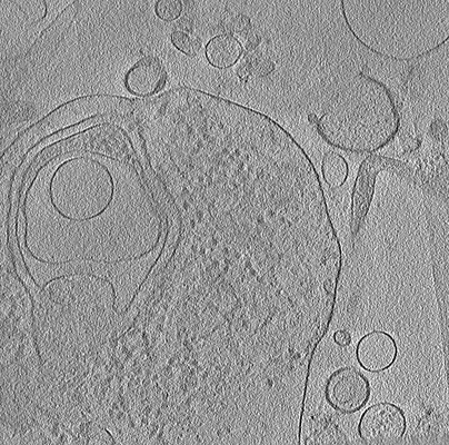
 ダウンロードとリンク
 emd_7881.png
 http://ftp.pdbj.org/pub/emdb/structures/EMD-7881


 試料の構成要素
 Homo sapiens (ヒト)
 解析
解析 電子顕微鏡法
 FIELD EMISSION GUN
