 Movie
Movie Controller


+ Open data

- Basic information
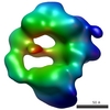
| Entry | Database: EMDB / ID: EMD-7827 | |||||||||
|---|---|---|---|---|---|---|---|---|---|---|
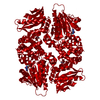
| Title | AprA Methyltransferase 1 - GNAT - Methyltransferase 2 tridomain | |||||||||
 Map data Map data | AprA Methyltransferase 1 - GNAT - Methyltransferase 2 tridomain | |||||||||
 Sample Sample |
| |||||||||
| Biological species |  Moorea bouillonii PNG5-198 (bacteria) Moorea bouillonii PNG5-198 (bacteria) | |||||||||
| Method | single particle reconstruction / negative staining / Resolution: 26.0 Å | |||||||||
 Authors Authors | Su M / Skiba MA / Smith JL | |||||||||
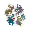
 Citation Citation | Journal: ACS Chem Biol / Year: 2018 Title: Biosynthesis of t-Butyl in Apratoxin A: Functional Analysis and Architecture of a PKS Loading Module. Authors: Meredith A Skiba / Andrew P Sikkema / Nathan A Moss / Andrew N Lowell / Min Su / Rebecca M Sturgis / Lena Gerwick / William H Gerwick / David H Sherman / Janet L Smith /  Abstract: The unusual feature of a t-butyl group is found in several marine-derived natural products including apratoxin A, a Sec61 inhibitor produced by the cyanobacterium Moorea bouillonii PNG 5-198. Here, ...The unusual feature of a t-butyl group is found in several marine-derived natural products including apratoxin A, a Sec61 inhibitor produced by the cyanobacterium Moorea bouillonii PNG 5-198. Here, we determine that the apratoxin A t-butyl group is formed as a pivaloyl acyl carrier protein (ACP) by AprA, the polyketide synthase (PKS) loading module of the apratoxin A biosynthetic pathway. AprA contains an inactive "pseudo" GCN5-related N-acetyltransferase domain (ΨGNAT) flanked by two methyltransferase domains (MT1 and MT2) that differ distinctly in sequence. Structural, biochemical, and precursor incorporation studies reveal that MT2 catalyzes unusually coupled decarboxylation and methylation reactions to transform dimethylmalonyl-ACP, the product of MT1, to pivaloyl-ACP. Further, pivaloyl-ACP synthesis is primed by the fatty acid synthase malonyl acyltransferase (FabD), which compensates for the ΨGNAT and provides the initial acyl-transfer step to form AprA malonyl-ACP. Additionally, images of AprA from negative stain electron microscopy reveal multiple conformations that may facilitate the individual catalytic steps of the multienzyme module. | |||||||||
| History |
|
- Structure visualization
| Movie |
 Movie viewer Movie viewer |
|---|---|
| Structure viewer | EM map: SurfViewMolmilJmol/JSmol |
| Supplemental images |
 UCSF Chimera
- Downloads & links
-EMDB archive
| Map data | emd_7827.map.gz | 1.5 MB | EMDB map data format | |
|---|---|---|---|---|
| Header (meta data) | emd-7827-v30.xmlemd-7827.xml | 10.5 KB 10.5 KB | Display Display | EMDB header |
| Images |  emd_7827.png emd_7827.png | 42.2 KB | ||
| Archive directory |  http://ftp.pdbj.org/pub/emdb/structures/EMD-7827ftp://ftp.pdbj.org/pub/emdb/structures/EMD-7827 http://ftp.pdbj.org/pub/emdb/structures/EMD-7827ftp://ftp.pdbj.org/pub/emdb/structures/EMD-7827 | HTTPS FTP |
-Related structure data


-Links
| EMDB pages | EMDB (EBI/PDBe) / EMDataResource |
|---|
-Map
| File | Download / File: emd_7827.map.gz / Format: CCP4 / Size: 2.6 MB / Type: IMAGE STORED AS FLOATING POINT NUMBER (4 BYTES) | ||||||||||||||||||||||||||||||||||||||||||||||||||||||||||||||||||||
|---|---|---|---|---|---|---|---|---|---|---|---|---|---|---|---|---|---|---|---|---|---|---|---|---|---|---|---|---|---|---|---|---|---|---|---|---|---|---|---|---|---|---|---|---|---|---|---|---|---|---|---|---|---|---|---|---|---|---|---|---|---|---|---|---|---|---|---|---|---|
| Annotation | AprA Methyltransferase 1 - GNAT - Methyltransferase 2 tridomain | ||||||||||||||||||||||||||||||||||||||||||||||||||||||||||||||||||||
| Projections & slices | Image control
Images are generated by Spider. | ||||||||||||||||||||||||||||||||||||||||||||||||||||||||||||||||||||
| Voxel size | X=Y=Z: 4.32 Å | ||||||||||||||||||||||||||||||||||||||||||||||||||||||||||||||||||||
| Density |
| ||||||||||||||||||||||||||||||||||||||||||||||||||||||||||||||||||||
| Symmetry | Space group: 1 | ||||||||||||||||||||||||||||||||||||||||||||||||||||||||||||||||||||
| Details | EMDB XML:
CCP4 map header:
| ||||||||||||||||||||||||||||||||||||||||||||||||||||||||||||||||||||
 Z (Sec.)
 Y (Row.)
 X (Col.)


-Supplemental data
- Sample components
-Entire : AprA Polyketide Synthase
| Entire | Name: AprA Polyketide Synthase |
|---|---|
| Components |
|
-Supramolecule #1: AprA Polyketide Synthase
| Supramolecule | Name: AprA Polyketide Synthase / type: complex / ID: 1 / Parent: 0 / Macromolecule list: all |
|---|---|
| Source (natural) | Organism: Moorea bouillonii PNG5-198 (bacteria) |
| Recombinant expression | Organism: |
-Macromolecule #1: AprA
| Macromolecule | Name: AprA / type: protein_or_peptide / ID: 1 / Enantiomer: DEXTRO |
|---|---|
| Source (natural) | Organism: Moorea bouillonii PNG5-198 (bacteria) |
| Recombinant expression | Organism: |
| Sequence | String: MHHHHHHSSG VDLGTENLYF QSNALDKIN RYAHGFVAVP VICACSEAGV FELLSQKKSL KLEEIVEHLA ANSG HLMVA MRLLESLSFL YRSQAEEYIL TEQSQQHQII PKALMSLYKY PFELY LKGE VETGISNWIN CSSRRWDTEN SLLSDLLDGV LLIPLLLELK ...String: MHHHHHHSSG VDLGTENLYF QSNALDKIN RYAHGFVAVP VICACSEAGV FELLSQKKSL KLEEIVEHLA ANSG HLMVA MRLLESLSFL YRSQAEEYIL TEQSQQHQII PKALMSLYKY PFELY LKGE VETGISNWIN CSSRRWDTEN SLLSDLLDGV LLIPLLLELK KQNLLD ESK KIFNTLTNSL KQELSTLFIN LGWAEEKTEG LYLTDIGRFM RDRSLNL GT TASYAPMLLQ MKELLFGNPQ RVFQRNKTEK ERHVNRTLNV VASGFQHE K FFADTDKIII SIFNQQPIEE QPSYIVDMGC GDGTLLKRIY KIIKQFSAR GKVLTEYPII MVGVDYNQEA LDVTDKNLVD IPHLVIPGDI GAPEKLLEQL KAQGIEPEK VLHIRSFLDH DRPFIAPKNT EIAQARSQLD YQVVDVDREG K LIPPHIAV QSLVEHLERW SSIITRHGLL LLEVHSLTPA VVKKYIDESE SL HFDAYHA FSMQHLVEAD VFLMAAAEVG LFSRKEAFRK YPKTLPLTRI TVN HFEKRK YQIRYATVND IPNLLKCATF NQPVNEPFFQ VLLKQTPTAH LLLE YQGEL VAAIFTETKN SNEVLGIREF LVRTSVENWQ VLAKDLLEFV EQWGV VKPG IKEIEGLLKY HEAISNFQKS KWYQSSVLNK KLIEKITLHE LATLEL CNL MAPEYELEAF AARWLLRVFQ DMGVFLREGE SYQESELVSQ LNISPRY QR LLGALLQILH KRGILKIEKD RVFTLARCKT FALENISSEV SAFYDYFS E KYPAHLSWLT VVKRCLEKYP LILRGEVDVN EVVFTDGDME LFAGLFLGH RVADYFNELL ADGVCWEVEQ RLLEEKRAQP IRILEIGAGT GGVTGILLEK LASHAEQIE FWFTDISSVF TRYGESKFKQ FPWVKYQTFD IEKSLDAQGI K SESFDVVI ANNVLHNTKL IHQTLNNSNS LLNTGGLLAL LEFTQPIDIL LY FGGLLQG FWLFEDPEYR LEVGCLLSIP LWQKVLSDCG FDEIIPLGLP CEM HALSKA RESVIFARKH QVQEKTFSEK IKQNLTEN |
-Experimental details
-Structure determination
| Method | negative staining |
|---|---|
 Processing Processing | single particle reconstruction |
| Aggregation state | particle |
-Sample preparation
| Buffer | pH: 7 |
|---|---|
| Staining | Type: NEGATIVE / Material: uranyl formate |
- Electron microscopy
| Microscope | FEI TECNAI 12 |
|---|---|
| Image recording | Film or detector model: GATAN ULTRASCAN 4000 (4k x 4k) / Average electron dose: 30.0 e/Å2 |
| Electron beam | Acceleration voltage: 120 kV / Electron source: LAB6 |
| Electron optics | Illumination mode: FLOOD BEAM / Imaging mode: BRIGHT FIELD |
-Image processing
| CTF correction | Software - Name: RELION (ver. 1.4) |
|---|---|
| Final reconstruction | Applied symmetry - Point group: C2 (2 fold cyclic) / Resolution.type: BY AUTHOR / Resolution: 26.0 Å / Resolution method: FSC 0.143 CUT-OFF / Software - Name: RELION (ver. 1.4) / Number images used: 4000 |
| Initial angle assignment | Type: COMMON LINE / Software - Name: EMAN2 (ver. 2.1) |
| Final angle assignment | Type: ANGULAR RECONSTITUTION / Software - Name: RELION (ver. 1.4) |
