 ムービー
ムービー コントローラー


+ データを開く

- 基本情報
基本情報
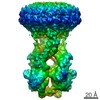
| 登録情報 | データベース: EMDB / ID: EMD-7802 | |||||||||
|---|---|---|---|---|---|---|---|---|---|---|
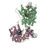
| タイトル | Cryo-EM map of F.graminearum Mitochondrial Calcium Uniporter in lipid nanodisc - type 2 | |||||||||
 マップデータ マップデータ | Cryo-EM map of FgMCU in nanodisc -type 2 filtered to 4.8A with -240 bfactor | |||||||||
 試料 試料 |
| |||||||||
| 生物種 |  Fusarium graminearum (ムギノアカカビ病菌) Fusarium graminearum (ムギノアカカビ病菌) | |||||||||
| 手法 | 単粒子再構成法 / クライオ電子顕微鏡法 / 解像度: 4.9 Å | |||||||||
 データ登録者 データ登録者 | Orlando BJ / Liao M | |||||||||
 引用 引用 | ジャーナル: Nature / 年: 2018 タイトル: X-ray and cryo-EM structures of the mitochondrial calcium uniporter. 著者: Chao Fan / Minrui Fan / Benjamin J Orlando / Nathan M Fastman / Jinru Zhang / Yan Xu / Melissa G Chambers / Xiaofang Xu / Kay Perry / Maofu Liao / Liang Feng /   要旨: Mitochondrial calcium uptake is critical for regulating ATP production, intracellular calcium signalling, and cell death. This uptake is mediated by a highly selective calcium channel called the ...Mitochondrial calcium uptake is critical for regulating ATP production, intracellular calcium signalling, and cell death. This uptake is mediated by a highly selective calcium channel called the mitochondrial calcium uniporter (MCU). Here, we determined the structures of the pore-forming MCU proteins from two fungi by X-ray crystallography and single-particle cryo-electron microscopy. The stoichiometry, overall architecture, and individual subunit structure differed markedly from those described in the recent nuclear magnetic resonance structure of Caenorhabditis elegans MCU. We observed a dimer-of-dimer architecture across species and chemical environments, which was corroborated by biochemical experiments. Structural analyses and functional characterization uncovered the roles of key residues in the pore. These results reveal a new ion channel architecture, provide insights into calcium coordination, selectivity and conduction, and establish a structural framework for understanding the mechanism of mitochondrial calcium uniporter function. | |||||||||
| 履歴 |
|
- 構造の表示
構造の表示
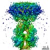
| ムービー |
 ムービービューア ムービービューア |
|---|---|
| 構造ビューア | EMマップ: SurfViewMolmilJmol/JSmol |
| 添付画像 |

- ダウンロードとリンク
-EMDBアーカイブ
| マップデータ | emd_7802.map.gz | 24.6 MB | EMDBマップデータ形式 | |
|---|---|---|---|---|
| ヘッダ (付随情報) | emd-7802-v30.xmlemd-7802.xml | 13.4 KB 13.4 KB | 表示 表示 | EMDBヘッダ |
| FSC (解像度算出) | emd_7802_fsc.xml | 8 KB | 表示 | FSCデータファイル |
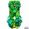
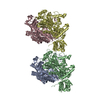
| 画像 |  emd_7802.png emd_7802.png | 62.7 KB | ||
| その他 | emd_7802_additional.map.gz | 19.7 MB | ||
| アーカイブディレクトリ |  http://ftp.pdbj.org/pub/emdb/structures/EMD-7802ftp://ftp.pdbj.org/pub/emdb/structures/EMD-7802 http://ftp.pdbj.org/pub/emdb/structures/EMD-7802ftp://ftp.pdbj.org/pub/emdb/structures/EMD-7802 | HTTPS FTP |
-関連構造データ


-リンク
| EMDBのページ | EMDB (EBI/PDBe) / EMDataResource |
|---|
-マップ
| ファイル | ダウンロード / ファイル: emd_7802.map.gz / 形式: CCP4 / 大きさ: 27 MB / タイプ: IMAGE STORED AS FLOATING POINT NUMBER (4 BYTES) | ||||||||||||||||||||||||||||||||||||||||||||||||||||||||||||
|---|---|---|---|---|---|---|---|---|---|---|---|---|---|---|---|---|---|---|---|---|---|---|---|---|---|---|---|---|---|---|---|---|---|---|---|---|---|---|---|---|---|---|---|---|---|---|---|---|---|---|---|---|---|---|---|---|---|---|---|---|---|
| 注釈 | Cryo-EM map of FgMCU in nanodisc -type 2 filtered to 4.8A with -240 bfactor | ||||||||||||||||||||||||||||||||||||||||||||||||||||||||||||
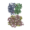
| 投影像・断面図 | 画像のコントロール
画像は Spider により作成 | ||||||||||||||||||||||||||||||||||||||||||||||||||||||||||||
| ボクセルのサイズ | X=Y=Z: 1.169 Å | ||||||||||||||||||||||||||||||||||||||||||||||||||||||||||||
| 密度 |
| ||||||||||||||||||||||||||||||||||||||||||||||||||||||||||||
| 対称性 | 空間群: 1 | ||||||||||||||||||||||||||||||||||||||||||||||||||||||||||||
| 詳細 | EMDB XML:
CCP4マップ ヘッダ情報:
| ||||||||||||||||||||||||||||||||||||||||||||||||||||||||||||
 Z (Sec.)
 Y (Row.)
 X (Col.)


-添付データ
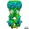
-追加マップ: unfiltered cryo-EM map of FgMCU in nanodisc -type 2
| ファイル | emd_7802_additional.map | ||||||||||||
|---|---|---|---|---|---|---|---|---|---|---|---|---|---|
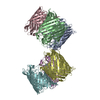
| 注釈 | unfiltered cryo-EM map of FgMCU in nanodisc -type 2 | ||||||||||||
| 投影像・断面図 |
| ||||||||||||
| 密度ヒストグラム |


- 試料の構成要素
-全体 : Fusarium graminearum Mitochondrial Calcium Uniporter in lipid nan...
| 全体 | 名称: Fusarium graminearum Mitochondrial Calcium Uniporter in lipid nanodiscs - type 2 |
|---|---|
| 要素 |
|
-超分子 #1: Fusarium graminearum Mitochondrial Calcium Uniporter in lipid nan...
| 超分子 | 名称: Fusarium graminearum Mitochondrial Calcium Uniporter in lipid nanodiscs - type 2 タイプ: complex / ID: 1 / 親要素: 0 |
|---|---|
| 由来(天然) | 生物種: Fusarium graminearum (ムギノアカカビ病菌) |
| 組換発現 | 生物種:  |
-実験情報
-構造解析
| 手法 | クライオ電子顕微鏡法 |
|---|---|
 解析 解析 | 単粒子再構成法 |
| 試料の集合状態 | particle |
-試料調製
| 濃度 | 5 mg/mL | |||||||||||||||
|---|---|---|---|---|---|---|---|---|---|---|---|---|---|---|---|---|
| 緩衝液 | pH: 8 構成要素:
| |||||||||||||||
| グリッド | モデル: Quantifoil R1.2/1.3 / 前処理 - タイプ: GLOW DISCHARGE / 詳細: 15mA in Pelco EasyGlow | |||||||||||||||
| 凍結 | 凍結剤: ETHANE / チャンバー内湿度: 92 % / 装置: GATAN CRYOPLUNGE 3 |
- 電子顕微鏡法
| 顕微鏡 | FEI TITAN KRIOS |
|---|---|
| 撮影 | フィルム・検出器のモデル: GATAN K2 SUMMIT (4k x 4k) 検出モード: SUPER-RESOLUTION / 平均電子線量: 46.0 e/Å2 |
| 電子線 | 加速電圧: 300 kV / 電子線源:  FIELD EMISSION GUN FIELD EMISSION GUN |
| 電子光学系 | 照射モード: FLOOD BEAM / 撮影モード: BRIGHT FIELD / Cs: 2.7 mm |
| 実験機器 |  モデル: Titan Krios / 画像提供: FEI Company |

