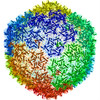
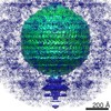
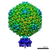
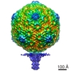
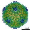
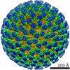
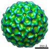
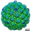
ジャーナル: Biophys J / 年: 2018 タイトル: Cryo-EM Elucidation of the Structure of Bacteriophage P22 Virions after Genome Release. 著者: Reginald McNulty / Giovanni Cardone / Eddie B Gilcrease / Timothy S Baker / Sherwood R Casjens / John E Johnson / 要旨: Genome ejection proteins are required to facilitate transport of bacteriophage P22 double-stranded DNA safely through membranes of Salmonella. The structures and locations of all proteins in the ...Genome ejection proteins are required to facilitate transport of bacteriophage P22 double-stranded DNA safely through membranes of Salmonella. The structures and locations of all proteins in the context of the mature virion are known, with the exception of three ejection proteins. Furthermore, the changes that occur to the proteins residing in the mature virion upon DNA release are not fully understood. We used cryogenic electron microscopy to obtain what is, to our knowledge, the first asymmetric reconstruction of mature bacteriophage P22 after double-stranded DNA has been extruded from the capsid-a state representative of one step during viral infection. Results of icosahedral and asymmetric reconstructions at estimated resolutions of 7.8 and 12.5 Å resolutions, respectively, are presented. The reconstruction shows tube-like protein density extending from the center of the tail assembly. The portal protein does not revert to the more contracted, procapsid state, but instead maintains an extended and splayed barrel structure. These structural details contribute to our understanding of the molecular mechanism of P22 phage infection and also set the foundation for future exploitation serving engineering purposes.
 ムービー
ムービー コントローラー


 データを開く
 基本情報
基本情報 マップデータ
 試料
試料 Enterobacteria phage P22 (ファージ)
 データ登録者
 引用
引用
 構造の表示
構造の表示 ムービービューア
 ダウンロードとリンク
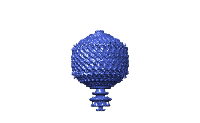 emd_7316.png
 http://ftp.pdbj.org/pub/emdb/structures/EMD-7316
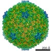

 Z (Sec.)
 Y (Row.)
 X (Col.)


 試料の構成要素
 Salmonella enterica subsp. enterica serovar Typhimurium (サルモネラ菌)
 解析
解析 電子顕微鏡法
 FIELD EMISSION GUN