[English] 日本語
 Yorodumi
- EMDB-7306: Structure of PRC2 bound to a H3K27me3/WT hetero-dinucleosome subs... -
+ Open data
- Basic information
| Entry | Database: EMDB / ID: EMD-7306 | |||||||||
|---|---|---|---|---|---|---|---|---|---|---|
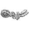
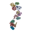
| Title | Structure of PRC2 bound to a H3K27me3/WT hetero-dinucleosome substrate with a 35 bp DNA linker | |||||||||
 Map data Map data | Complete Map of the PRC2 catalyitic lobe bound to a hetero-dinucleosome | |||||||||
 Sample Sample |
| |||||||||
| Biological species |  Homo sapiens (human) Homo sapiens (human) | |||||||||
| Method | single particle reconstruction / cryo EM / Resolution: 6.2 Å | |||||||||
 Authors Authors | Poepsel S / Kasinath V / Nogales E | |||||||||
 Citation Citation |  Journal: Nat Struct Mol Biol / Year: 2018 Journal: Nat Struct Mol Biol / Year: 2018Title: Cryo-EM structures of PRC2 simultaneously engaged with two functionally distinct nucleosomes. Authors: Simon Poepsel / Vignesh Kasinath / Eva Nogales /  Abstract: Epigenetic regulation is mediated by protein complexes that couple recognition of chromatin marks to activity or recruitment of chromatin-modifying enzymes. Polycomb repressive complex 2 (PRC2), a ...Epigenetic regulation is mediated by protein complexes that couple recognition of chromatin marks to activity or recruitment of chromatin-modifying enzymes. Polycomb repressive complex 2 (PRC2), a gene silencer that methylates lysine 27 of histone H3, is stimulated upon recognition of its own catalytic product and has been shown to be more active on dinucleosomes than H3 tails or single nucleosomes. These properties probably facilitate local H3K27me2/3 spreading, causing heterochromatin formation and gene repression. Here, cryo-EM reconstructions of human PRC2 bound to bifunctional dinucleosomes show how a single PRC2, via interactions with nucleosomal DNA, positions the H3 tails of the activating and substrate nucleosome to interact with the EED subunit and the SET domain of EZH2, respectively. We show how the geometry of the PRC2-DNA interactions allows PRC2 to accommodate varying lengths of the linker DNA between nucleosomes. Our structures illustrate how an epigenetic regulator engages with a complex chromatin substrate. | |||||||||
| History |
|
- Structure visualization
| Movie |
 Movie viewer Movie viewer |
|---|---|
| Structure viewer | EM map:  SurfView SurfView Molmil Molmil Jmol/JSmol Jmol/JSmol |
| Supplemental images |
- Downloads & links
-EMDB archive
| Map data |  emd_7306.map.gz emd_7306.map.gz | 166.5 MB |  EMDB map data format EMDB map data format | |
|---|---|---|---|---|
| Header (meta data) |  emd-7306-v30.xml emd-7306-v30.xml emd-7306.xml emd-7306.xml | 15.3 KB 15.3 KB | Display Display |  EMDB header EMDB header |
| FSC (resolution estimation) |  emd_7306_fsc.xml emd_7306_fsc.xml | 12.5 KB | Display |  FSC data file FSC data file |
| Images |  emd_7306.png emd_7306.png | 35.7 KB | ||
| Others |  emd_7306_half_map_1.map.gz emd_7306_half_map_1.map.gz emd_7306_half_map_2.map.gz emd_7306_half_map_2.map.gz | 140.9 MB 140.9 MB | ||
| Archive directory |  http://ftp.pdbj.org/pub/emdb/structures/EMD-7306 http://ftp.pdbj.org/pub/emdb/structures/EMD-7306 ftp://ftp.pdbj.org/pub/emdb/structures/EMD-7306 ftp://ftp.pdbj.org/pub/emdb/structures/EMD-7306 | HTTPS FTP |
-Validation report
| Summary document |  emd_7306_validation.pdf.gz emd_7306_validation.pdf.gz | 458.4 KB | Display |  EMDB validaton report EMDB validaton report |
|---|---|---|---|---|
| Full document |  emd_7306_full_validation.pdf.gz emd_7306_full_validation.pdf.gz | 457.9 KB | Display | |
| Data in XML |  emd_7306_validation.xml.gz emd_7306_validation.xml.gz | 18.1 KB | Display | |
| Arichive directory |  https://ftp.pdbj.org/pub/emdb/validation_reports/EMD-7306 https://ftp.pdbj.org/pub/emdb/validation_reports/EMD-7306 ftp://ftp.pdbj.org/pub/emdb/validation_reports/EMD-7306 ftp://ftp.pdbj.org/pub/emdb/validation_reports/EMD-7306 | HTTPS FTP |
-Related structure data
| Related structure data |  7307C  7308C  7309C  7310C  7311C  7312C  7313C C: citing same article ( |
|---|---|
| Similar structure data |
- Links
Links
| EMDB pages |  EMDB (EBI/PDBe) / EMDB (EBI/PDBe) /  EMDataResource EMDataResource |
|---|
- Map
Map
| File |  Download / File: emd_7306.map.gz / Format: CCP4 / Size: 178 MB / Type: IMAGE STORED AS FLOATING POINT NUMBER (4 BYTES) Download / File: emd_7306.map.gz / Format: CCP4 / Size: 178 MB / Type: IMAGE STORED AS FLOATING POINT NUMBER (4 BYTES) | ||||||||||||||||||||||||||||||||||||||||||||||||||||||||||||||||||||
|---|---|---|---|---|---|---|---|---|---|---|---|---|---|---|---|---|---|---|---|---|---|---|---|---|---|---|---|---|---|---|---|---|---|---|---|---|---|---|---|---|---|---|---|---|---|---|---|---|---|---|---|---|---|---|---|---|---|---|---|---|---|---|---|---|---|---|---|---|---|
| Annotation | Complete Map of the PRC2 catalyitic lobe bound to a hetero-dinucleosome | ||||||||||||||||||||||||||||||||||||||||||||||||||||||||||||||||||||
| Projections & slices | Image control
Images are generated by Spider. | ||||||||||||||||||||||||||||||||||||||||||||||||||||||||||||||||||||
| Voxel size | X=Y=Z: 1.32 Å | ||||||||||||||||||||||||||||||||||||||||||||||||||||||||||||||||||||
| Density |
| ||||||||||||||||||||||||||||||||||||||||||||||||||||||||||||||||||||
| Symmetry | Space group: 1 | ||||||||||||||||||||||||||||||||||||||||||||||||||||||||||||||||||||
| Details | EMDB XML:
CCP4 map header:
| ||||||||||||||||||||||||||||||||||||||||||||||||||||||||||||||||||||
-Supplemental data
-Half map: half map 1
| File | emd_7306_half_map_1.map | ||||||||||||
|---|---|---|---|---|---|---|---|---|---|---|---|---|---|
| Annotation | half map 1 | ||||||||||||
| Projections & Slices |
| ||||||||||||
| Density Histograms |
-Half map: half map 2
| File | emd_7306_half_map_2.map | ||||||||||||
|---|---|---|---|---|---|---|---|---|---|---|---|---|---|
| Annotation | half map 2 | ||||||||||||
| Projections & Slices |
| ||||||||||||
| Density Histograms |
- Sample components
-Entire : Human Polycomb Repressive Complex 2 (PRC2) in complex with a hete...
| Entire | Name: Human Polycomb Repressive Complex 2 (PRC2) in complex with a heterodinucleosome composed of an unmodified nucleosome and a nucleosome carrying an H3K27 pseudo-trimethyl mark |
|---|---|
| Components |
|
-Supramolecule #1: Human Polycomb Repressive Complex 2 (PRC2) in complex with a hete...
| Supramolecule | Name: Human Polycomb Repressive Complex 2 (PRC2) in complex with a heterodinucleosome composed of an unmodified nucleosome and a nucleosome carrying an H3K27 pseudo-trimethyl mark type: complex / ID: 1 / Parent: 0 / Macromolecule list: all |
|---|---|
| Source (natural) | Organism:  Homo sapiens (human) Homo sapiens (human) |
| Recombinant expression | Organism:  Trichoplusia ni (cabbage looper) Trichoplusia ni (cabbage looper) |
-Macromolecule #1: EZH2
| Macromolecule | Name: EZH2 / type: protein_or_peptide / ID: 1 / Enantiomer: LEVO |
|---|---|
| Sequence | String: MGQTGKKSEK GPVCWRKRVK SEYMRLRQLK RFRRADEVKS MFSSNRQKIL ERTEILNQEW KQRRIQPVH ILTSVSSLRG TRECSVTSDL DFPTQVIPLK TLNAVASVPI MYSWSPLQQN F MVEDETVL HNIPYMGDEV LDQDGTFIEE LIKNYDGKVH GDRECGFIND ...String: MGQTGKKSEK GPVCWRKRVK SEYMRLRQLK RFRRADEVKS MFSSNRQKIL ERTEILNQEW KQRRIQPVH ILTSVSSLRG TRECSVTSDL DFPTQVIPLK TLNAVASVPI MYSWSPLQQN F MVEDETVL HNIPYMGDEV LDQDGTFIEE LIKNYDGKVH GDRECGFIND EIFVELVNAL GQ YNDDDDD DDGDDPEERE EKQKDLEDHR DDKESRPPRK FPSDKIFEAI SSMFPDKGTA EEL KEKYKE LTEQQLPGAL PPECTPNIDG PNAKSVQREQ SLHSFHTLFC RRCFKYDCFL HPFH ATPNT YKRKNTETAL DNKPCGPQCY QHLEGAKEFA AALTAERIKT PPKRPGGRRR GRLPN NSSR PSTPTINVLE SKDTDSDREA GTETGGENND KEEEEKKDET SSSSEANSRC QTPIKM KPN IEPPENVEWS GAEASMFRVL IGTYYDNFCA IARLIGTKTC RQVYEFRVKE SSIIAPA PA EDVDTPPRKK KRKHRLWAAH CRKIQLKKDG SSNHVYNYQP CDHPRQPCDS SCPCVIAQ N FCEKFCQCSS ECQNRFPGCR CKAQCNTKQC PCYLAVRECD PDLCLTCGAA DHWDSKNVS CKNCSIQRGS KKHLLLAPSD VAGWGIFIKD PVQKNEFISE YCGEIISQDE ADRRGKVYDK YMCSFLFNL NNDFVVDATR KGNKIRFANH SVNPNCYAKV MMVNGDHRIG IFAKRAIQTG E ELFFDYRY SQADALKYVG IEREMEIP |
-Experimental details
-Structure determination
| Method | cryo EM |
|---|---|
 Processing Processing | single particle reconstruction |
| Aggregation state | particle |
- Sample preparation
| Concentration | 1 mg/mL |
|---|---|
| Buffer | pH: 7.9 |
| Grid | Model: Quantifoil R1.2/1.3 / Material: COPPER / Mesh: 400 |
| Vitrification | Cryogen name: ETHANE / Chamber humidity: 100 % / Chamber temperature: 281 K / Instrument: FEI VITROBOT MARK IV |
- Electron microscopy
| Microscope | FEI TITAN |
|---|---|
| Image recording | Film or detector model: GATAN K2 SUMMIT (4k x 4k) / Detector mode: COUNTING / Number grids imaged: 3 / Number real images: 4600 / Average exposure time: 8.7 sec. / Average electron dose: 40.0 e/Å2 |
| Electron beam | Acceleration voltage: 300 kV / Electron source:  FIELD EMISSION GUN FIELD EMISSION GUN |
| Electron optics | Illumination mode: FLOOD BEAM / Imaging mode: BRIGHT FIELD |
 Movie
Movie Controller


 Z (Sec.)
 Y (Row.)
 X (Col.)


