 Movie
Movie Controller

[English] 日本語
 Yorodumi
- EMDB-7311: Structure of PRC2 bound to a H3K27me3/WT hetero-dinucleosome subs... -
+ Open data

- Basic information
| Entry | Database: EMDB / ID: EMD-7311 | |||||||||
|---|---|---|---|---|---|---|---|---|---|---|
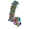
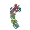
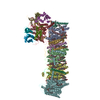
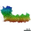
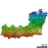
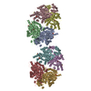
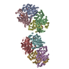
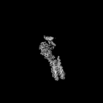
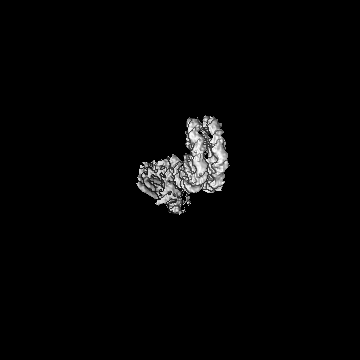
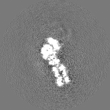
| Title | Structure of PRC2 bound to a H3K27me3/WT hetero-dinucleosome substrate with a 35 bp DNA linker. Masked refinement of PRC2-substrate nucleosome subcomplex. | |||||||||
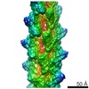
 Map data Map data | Structure of human PRC2 bound to a hetero-dinucleosome substrate with 35 bp linker DNA. Refinement after signal subtraction of the modified nucleosome. | |||||||||
 Sample Sample |
| |||||||||
| Biological species |  Homo sapiens (human) Homo sapiens (human) | |||||||||
| Method | single particle reconstruction / cryo EM / Resolution: 4.9 Å | |||||||||
 Authors Authors | Poepsel S / Kasinath V / Nogales E | |||||||||
 Citation Citation | Journal: Nat Struct Mol Biol / Year: 2018 Title: Cryo-EM structures of PRC2 simultaneously engaged with two functionally distinct nucleosomes. Authors: Simon Poepsel / Vignesh Kasinath / Eva Nogales /  Abstract: Epigenetic regulation is mediated by protein complexes that couple recognition of chromatin marks to activity or recruitment of chromatin-modifying enzymes. Polycomb repressive complex 2 (PRC2), a ...Epigenetic regulation is mediated by protein complexes that couple recognition of chromatin marks to activity or recruitment of chromatin-modifying enzymes. Polycomb repressive complex 2 (PRC2), a gene silencer that methylates lysine 27 of histone H3, is stimulated upon recognition of its own catalytic product and has been shown to be more active on dinucleosomes than H3 tails or single nucleosomes. These properties probably facilitate local H3K27me2/3 spreading, causing heterochromatin formation and gene repression. Here, cryo-EM reconstructions of human PRC2 bound to bifunctional dinucleosomes show how a single PRC2, via interactions with nucleosomal DNA, positions the H3 tails of the activating and substrate nucleosome to interact with the EED subunit and the SET domain of EZH2, respectively. We show how the geometry of the PRC2-DNA interactions allows PRC2 to accommodate varying lengths of the linker DNA between nucleosomes. Our structures illustrate how an epigenetic regulator engages with a complex chromatin substrate. | |||||||||
| History |
|
- Structure visualization
| Movie |
 Movie viewer Movie viewer |
|---|---|
| Structure viewer | EM map: SurfViewMolmilJmol/JSmol |
| Supplemental images |

- Downloads & links
-EMDB archive
| Map data | emd_7311.map.gz | 166.6 MB | EMDB map data format | |
|---|---|---|---|---|
| Header (meta data) | emd-7311-v30.xmlemd-7311.xml | 9.9 KB 9.9 KB | Display Display | EMDB header |
| FSC (resolution estimation) | emd_7311_fsc.xml | 12.6 KB | Display | FSC data file |
| Images | 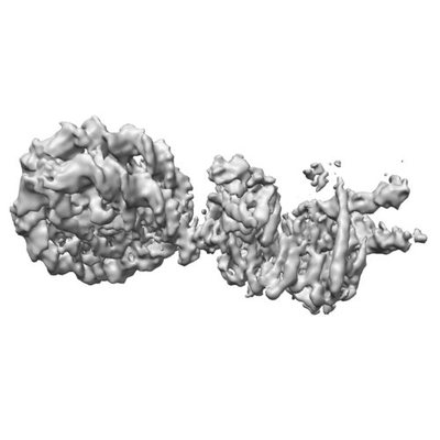 emd_7311.png emd_7311.png | 41 KB | ||
| Archive directory |  http://ftp.pdbj.org/pub/emdb/structures/EMD-7311ftp://ftp.pdbj.org/pub/emdb/structures/EMD-7311 http://ftp.pdbj.org/pub/emdb/structures/EMD-7311ftp://ftp.pdbj.org/pub/emdb/structures/EMD-7311 | HTTPS FTP |
-Related structure data
| Related structure data | 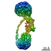 7306C 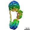 7307C 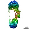 7308C 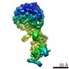 7309C 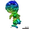 7310C 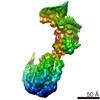 7312C 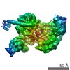 7313C C: citing same article ( |
|---|---|
| Similar structure data |
-Links
| EMDB pages | EMDB (EBI/PDBe) / EMDataResource |
|---|
-Map
| File | Download / File: emd_7311.map.gz / Format: CCP4 / Size: 178 MB / Type: IMAGE STORED AS FLOATING POINT NUMBER (4 BYTES) | ||||||||||||||||||||||||||||||||||||||||||||||||||||||||||||||||||||
|---|---|---|---|---|---|---|---|---|---|---|---|---|---|---|---|---|---|---|---|---|---|---|---|---|---|---|---|---|---|---|---|---|---|---|---|---|---|---|---|---|---|---|---|---|---|---|---|---|---|---|---|---|---|---|---|---|---|---|---|---|---|---|---|---|---|---|---|---|---|
| Annotation | Structure of human PRC2 bound to a hetero-dinucleosome substrate with 35 bp linker DNA. Refinement after signal subtraction of the modified nucleosome. | ||||||||||||||||||||||||||||||||||||||||||||||||||||||||||||||||||||
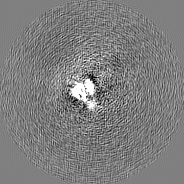
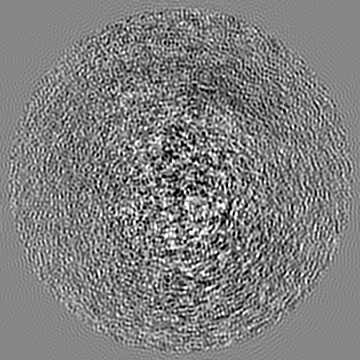
| Projections & slices | Image control
Images are generated by Spider. | ||||||||||||||||||||||||||||||||||||||||||||||||||||||||||||||||||||
| Voxel size | X=Y=Z: 1.32 Å | ||||||||||||||||||||||||||||||||||||||||||||||||||||||||||||||||||||
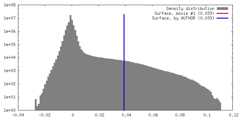
| Density |
| ||||||||||||||||||||||||||||||||||||||||||||||||||||||||||||||||||||
| Symmetry | Space group: 1 | ||||||||||||||||||||||||||||||||||||||||||||||||||||||||||||||||||||
| Details | EMDB XML:
CCP4 map header:
| ||||||||||||||||||||||||||||||||||||||||||||||||||||||||||||||||||||
 Z (Sec.)
 Y (Row.)
 X (Col.)


-Supplemental data
- Sample components
-Entire : Structure of human PRC2 bound to a hetero-dinucleosome substrate ...
| Entire | Name: Structure of human PRC2 bound to a hetero-dinucleosome substrate with 35 bp linker DNA. Refinement after signal subtraction to increase resolution of the substrate nucleosome - PRC2 interface. |
|---|---|
| Components |
|
-Supramolecule #1: Structure of human PRC2 bound to a hetero-dinucleosome substrate ...
| Supramolecule | Name: Structure of human PRC2 bound to a hetero-dinucleosome substrate with 35 bp linker DNA. Refinement after signal subtraction to increase resolution of the substrate nucleosome - PRC2 interface. type: complex / ID: 1 / Parent: 0 / Macromolecule list: #1 |
|---|---|
| Source (natural) | Organism: Homo sapiens (human) |
| Recombinant expression | Organism:  Trichoplusia ni (cabbage looper) Trichoplusia ni (cabbage looper) |
-Experimental details
-Structure determination
| Method | cryo EM |
|---|---|
 Processing Processing | single particle reconstruction |
| Aggregation state | particle |
-Sample preparation
| Concentration | 1 mg/mL |
|---|---|
| Buffer | pH: 7.9 |
| Vitrification | Cryogen name: ETHANE / Chamber humidity: 100 % / Chamber temperature: 281 K / Instrument: FEI VITROBOT MARK IV |
- Electron microscopy
| Microscope | FEI TITAN |
|---|---|
| Image recording | Film or detector model: GATAN K2 SUMMIT (4k x 4k) / Average electron dose: 40.0 e/Å2 |
| Electron beam | Acceleration voltage: 300 kV / Electron source:  FIELD EMISSION GUN FIELD EMISSION GUN |
| Electron optics | Illumination mode: FLOOD BEAM / Imaging mode: BRIGHT FIELD |
-Image processing
| Final reconstruction | Applied symmetry - Point group: C1 (asymmetric) / Resolution.type: BY AUTHOR / Resolution: 4.9 Å / Resolution method: FSC 0.143 CUT-OFF Details: Signal subtraction of the particle images was performed to exclude signal from the more flexible modified nucleosome from the reconstruction. Number images used: 93384 |
|---|---|
| Initial angle assignment | Type: PROJECTION MATCHING |
| Final angle assignment | Type: PROJECTION MATCHING |
| FSC plot (resolution estimation) | 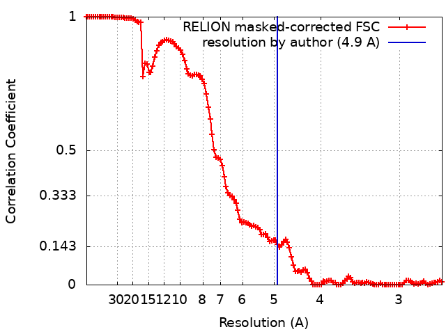 |
