Movie
Movie Controller

[English] 日本語
 Yorodumi
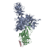
- EMDB-72215: Focused cryo-EM map of DDB1dB:CRBN:mezigdomide:SALL4(392-449; G416A) -
+ Open data

- Basic information
| Entry |  | |||||||||
|---|---|---|---|---|---|---|---|---|---|---|
| Title | Focused cryo-EM map of DDB1dB:CRBN:mezigdomide:SALL4(392-449; G416A) | |||||||||
 Map data Map data | Focused map, auto-sharpened by cryoSPARC | |||||||||
 Sample Sample |
| |||||||||
 Keywords Keywords | DDB1 / cereblon / mezigdomide / SALL4 / LIGASE | |||||||||
| Function / homology |  Function and homology information Function and homology informationPOU5F1 (OCT4), SOX2, NANOG activate genes related to proliferation / negative regulation of monoatomic ion transmembrane transport / Transcriptional regulation of pluripotent stem cells / positive regulation by virus of viral protein levels in host cell / spindle assembly involved in female meiosis / epigenetic programming in the zygotic pronuclei / UV-damage excision repair / embryonic limb morphogenesis / biological process involved in interaction with symbiont / inner cell mass cell proliferation ...POU5F1 (OCT4), SOX2, NANOG activate genes related to proliferation / negative regulation of monoatomic ion transmembrane transport / Transcriptional regulation of pluripotent stem cells / positive regulation by virus of viral protein levels in host cell / spindle assembly involved in female meiosis / epigenetic programming in the zygotic pronuclei / UV-damage excision repair / embryonic limb morphogenesis / biological process involved in interaction with symbiont / inner cell mass cell proliferation / regulation of mitotic cell cycle phase transition / ventricular septum development / WD40-repeat domain binding / Cul4A-RING E3 ubiquitin ligase complex / Cul4-RING E3 ubiquitin ligase complex / Cul4B-RING E3 ubiquitin ligase complex / ubiquitin ligase complex scaffold activity / negative regulation of reproductive process / negative regulation of developmental process / locomotory exploration behavior / cullin family protein binding / viral release from host cell / somatic stem cell population maintenance / positive regulation of Wnt signaling pathway / ectopic germ cell programmed cell death / negative regulation of protein-containing complex assembly / positive regulation of viral genome replication / heterochromatin / proteasomal protein catabolic process / positive regulation of gluconeogenesis / Regulation of PTEN gene transcription / nucleotide-excision repair / neural tube closure / positive regulation of protein-containing complex assembly / Recognition of DNA damage by PCNA-containing replication complex / DNA Damage Recognition in GG-NER / regulation of circadian rhythm / Dual Incision in GG-NER / Transcription-Coupled Nucleotide Excision Repair (TC-NER) / Formation of TC-NER Pre-Incision Complex / Formation of Incision Complex in GG-NER / Wnt signaling pathway / Dual incision in TC-NER / Gap-filling DNA repair synthesis and ligation in TC-NER / positive regulation of protein catabolic process / cellular response to UV / rhythmic process / site of double-strand break / Neddylation / ubiquitin-dependent protein catabolic process / protein-macromolecule adaptor activity / Potential therapeutics for SARS / proteasome-mediated ubiquitin-dependent protein catabolic process / damaged DNA binding / transmembrane transporter binding / DNA-binding transcription factor activity, RNA polymerase II-specific / chromosome, telomeric region / protein ubiquitination / DNA repair / intracellular membrane-bounded organelle / apoptotic process / DNA damage response / regulation of transcription by RNA polymerase II / negative regulation of apoptotic process / protein-containing complex binding / nucleolus / perinuclear region of cytoplasm / negative regulation of transcription by RNA polymerase II / positive regulation of transcription by RNA polymerase II / protein-containing complex / extracellular space / DNA binding / extracellular exosome / zinc ion binding / nucleoplasm / metal ion binding / nucleus / membrane / cytosol / cytoplasm Similarity search - Function | |||||||||
| Biological species |  Homo sapiens (human) Homo sapiens (human) | |||||||||
| Method | single particle reconstruction / cryo EM / Resolution: 2.93 Å | |||||||||
 Authors Authors | Park J / Hunkeler M / Roy Burman SS / Fischer ES | |||||||||
| Funding support |  United States, 1 items United States, 1 items
| |||||||||
 Citation Citation | Journal: Mol Cell / Year: 2025 Title: Expanding the druggable zinc-finger proteome defines properties of drug-induced degradation. Authors: Mikołaj Słabicki / Jiho Park / Radosław P Nowak / Shourya S Roy Burman / Jesse Pellman / Charles Zou / Hlib Razumkov / Jeannie Carreiro / Simran Rastogi / Anna Goldstein / Marek M Nagiec ...Authors: Mikołaj Słabicki / Jiho Park / Radosław P Nowak / Shourya S Roy Burman / Jesse Pellman / Charles Zou / Hlib Razumkov / Jeannie Carreiro / Simran Rastogi / Anna Goldstein / Marek M Nagiec / Katherine A Donovan / Jianwei Che / Moritz Hunkeler / Qixiang Geng / Chi-Lin Hsu / Megha Lakshminarayan / Chelsea Shu / Rebecca L Zon / Zuzanna Kozicka / Paul M C Park / Jonathan M Tsai / Hojong Yoon / Lyn H Jones / Adam S Sperling / Nathanael S Gray / Eric S Fischer / Benjamin L Ebert /  Abstract: Glutarimide analogs, such as thalidomide, redirect the E3 ubiquitin ligase CRL4 to induce degradation of certain zinc finger (ZF) proteins. Although the core structural motif recognized by CRBN has ...Glutarimide analogs, such as thalidomide, redirect the E3 ubiquitin ligase CRL4 to induce degradation of certain zinc finger (ZF) proteins. Although the core structural motif recognized by CRBN has been characterized, it does not fully explain substrate specificity. To explore the role of residues adjacent to this core motif, we constructed a comprehensive ZF reporter library of 9,097 reporters derived from 1,655 human ZF proteins and conducted a library-on-library screen with 29 glutarimide analogs to identify compounds that collectively degrade 38 ZF reporters. Cryo-electron microscopy and crystal structures of ZFs in complex with CRBN revealed the importance of interactions beyond the core ZF degron. We used systematic mutagenesis of ZFs and CRBN to identify modes of neosubstrate recruitment requiring distinct amino acids. Finally, we found subtle chemical variations in glutarimide analogs that alter target scope and selectivity, thus providing a roadmap for their rational design. | |||||||||
| History |
|
- Structure visualization
| Supplemental images |
|---|

- Downloads & links
-EMDB archive
| Map data | emd_72215.map.gz | 136.4 MB | EMDB map data format | |
|---|---|---|---|---|
| Header (meta data) | emd-72215-v30.xmlemd-72215.xml | 31.4 KB 31.4 KB | Display Display | EMDB header |
| Images |  emd_72215.png emd_72215.png | 71.9 KB | ||
| Masks | emd_72215_msk_1.map | 144.7 MB | Mask map | |
| Filedesc metadata | emd-72215.cif.gz | 8.1 KB | ||
| Others | emd_72215_additional_1.map.gzemd_72215_additional_2.map.gzemd_72215_half_map_1.map.gzemd_72215_half_map_2.map.gz | 72.1 MB 126.6 MB 134.5 MB 134.5 MB | ||
| Archive directory |  http://ftp.pdbj.org/pub/emdb/structures/EMD-72215ftp://ftp.pdbj.org/pub/emdb/structures/EMD-72215 http://ftp.pdbj.org/pub/emdb/structures/EMD-72215ftp://ftp.pdbj.org/pub/emdb/structures/EMD-72215 | HTTPS FTP |
-Validation report
| Summary document | emd_72215_validation.pdf.gz | 869.5 KB | Display | EMDB validaton report |
|---|---|---|---|---|
| Full document | emd_72215_full_validation.pdf.gz | 869 KB | Display | |
| Data in XML | emd_72215_validation.xml.gz | 14.2 KB | Display | |
| Data in CIF | emd_72215_validation.cif.gz | 16.9 KB | Display | |
| Arichive directory | https://ftp.pdbj.org/pub/emdb/validation_reports/EMD-72215ftp://ftp.pdbj.org/pub/emdb/validation_reports/EMD-72215 | HTTPS FTP |
-Related structure data
| Related structure data |  9e2uC  49893 C: citing same article ( |
|---|---|
| Similar structure data |

-Links
| EMDB pages | EMDB (EBI/PDBe) / EMDataResource |
|---|---|
| Related items in Molecule of the Month |


-Map
| File | Download / File: emd_72215.map.gz / Format: CCP4 / Size: 144.7 MB / Type: IMAGE STORED AS FLOATING POINT NUMBER (4 BYTES) | ||||||||||||||||||||||||||||||||||||
|---|---|---|---|---|---|---|---|---|---|---|---|---|---|---|---|---|---|---|---|---|---|---|---|---|---|---|---|---|---|---|---|---|---|---|---|---|---|
| Annotation | Focused map, auto-sharpened by cryoSPARC | ||||||||||||||||||||||||||||||||||||
| Projections & slices | Image control
Images are generated by Spider. | ||||||||||||||||||||||||||||||||||||
| Voxel size | X=Y=Z: 0.83 Å | ||||||||||||||||||||||||||||||||||||
| Density |
| ||||||||||||||||||||||||||||||||||||
| Symmetry | Space group: 1 | ||||||||||||||||||||||||||||||||||||
| Details | EMDB XML:
|
 Z (Sec.)
 Y (Row.)
 X (Col.)


-Supplemental data
-Mask #1
| File | emd_72215_msk_1.map | ||||||||||||
|---|---|---|---|---|---|---|---|---|---|---|---|---|---|
| Projections & Slices |
| ||||||||||||
| Density Histograms |


-Additional map: Focused map, unsharpened
| File | emd_72215_additional_1.map | ||||||||||||
|---|---|---|---|---|---|---|---|---|---|---|---|---|---|
| Annotation | Focused map, unsharpened | ||||||||||||
| Projections & Slices |
| ||||||||||||
| Density Histograms |


-Additional map: Focused map, post-processed with DeepEMhancer
| File | emd_72215_additional_2.map | ||||||||||||
|---|---|---|---|---|---|---|---|---|---|---|---|---|---|
| Annotation | Focused map, post-processed with DeepEMhancer | ||||||||||||
| Projections & Slices |
| ||||||||||||
| Density Histograms |


-Half map: Focused map, half-map A
| File | emd_72215_half_map_1.map | ||||||||||||
|---|---|---|---|---|---|---|---|---|---|---|---|---|---|
| Annotation | Focused map, half-map A | ||||||||||||
| Projections & Slices |
| ||||||||||||
| Density Histograms |


-Half map: Focused map, half-map A
| File | emd_72215_half_map_2.map | ||||||||||||
|---|---|---|---|---|---|---|---|---|---|---|---|---|---|
| Annotation | Focused map, half-map A | ||||||||||||
| Projections & Slices |
| ||||||||||||
| Density Histograms |


- Sample components
-Entire : Ternary complex of DDB1dB:CRBN:mezigdomide:SALL4(392-449;G416A)
| Entire | Name: Ternary complex of DDB1dB:CRBN:mezigdomide:SALL4(392-449;G416A) |
|---|---|
| Components |
|
-Supramolecule #1: Ternary complex of DDB1dB:CRBN:mezigdomide:SALL4(392-449;G416A)
| Supramolecule | Name: Ternary complex of DDB1dB:CRBN:mezigdomide:SALL4(392-449;G416A) type: complex / ID: 1 / Parent: 0 / Macromolecule list: all |
|---|---|
| Source (natural) | Organism: Homo sapiens (human) |
-Macromolecule #1: Sal-like protein 4
| Macromolecule | Name: Sal-like protein 4 / type: protein_or_peptide / ID: 1 / Details: StrepII-Avi-TEV-SALL4(392-449;G416A) / Enantiomer: LEVO |
|---|---|
| Source (natural) | Organism: Homo sapiens (human) |
| Recombinant expression | Organism:  Trichoplusia ni (cabbage looper) Trichoplusia ni (cabbage looper) |
| Sequence | String: MDWSHPQFEK SAVGLNDIFE AQKIEWHEGG GGSGENLYFQ GGGRGTDSSL QIHLRSHTGE RPFVCSVCAH RFTTKGNLKV HFHRHPQVK ANPQLFAEFQ DKV UniProtKB: Sal-like protein 4 |
-Macromolecule #2: DNA damage-binding protein 1
| Macromolecule | Name: DNA damage-binding protein 1 / type: protein_or_peptide / ID: 2 Details: His6-TEV-DDB1(1-1140), with residues 396-705 replaced with a GNGNSG linker,His6-TEV-DDB1(1-1140), with residues 396-705 replaced with a GNGNSG linker Enantiomer: LEVO |
|---|---|
| Source (natural) | Organism: Homo sapiens (human) |
| Recombinant expression | Organism: Trichoplusia ni (cabbage looper) |
| Sequence | String: MGSSHHHHHH SAVDENLYFQ GGGRMSYNYV VTAQKPTAVN GCVTGHFTSA EDLNLLIAKN TRLEIYVVTA EGLRPVKEVG MYGKIAVME LFRPKGESKD LLFILTAKYN ACILEYKQSG ESIDIITRAH GNVQDRIGRP SETGIIGIID PECRMIGLRL Y DGLFKVIP ...String: MGSSHHHHHH SAVDENLYFQ GGGRMSYNYV VTAQKPTAVN GCVTGHFTSA EDLNLLIAKN TRLEIYVVTA EGLRPVKEVG MYGKIAVME LFRPKGESKD LLFILTAKYN ACILEYKQSG ESIDIITRAH GNVQDRIGRP SETGIIGIID PECRMIGLRL Y DGLFKVIP LDRDNKELKA FNIRLEELHV IDVKFLYGCQ APTICFVYQD PQGRHVKTYE VSLREKEFNK GPWKQENVEA EA SMVIAVP EPFGGAIIIG QESITYHNGD KYLAIAPPII KQSTIVCHNR VDPNGSRYLL GDMEGRLFML LLEKEEQMDG TVT LKDLRV ELLGETSIAE CLTYLDNGVV FVGSRLGDSQ LVKLNVDSNE QGSYVVAMET FTNLGPIVDM CVVDLERQGQ GQLV TCSGA FKEGSLRIIR NGIGGNGNSG EIQKLHIRTV PLYESPRKIC YQEVSQCFGV LSSRIEVQDT SGGTTALRPS ASTQA LSSS VSSSKLFSSS TAPHETSFGE EVEVHNLLII DQHTFEVLHA HQFLQNEYAL SLVSCKLGKD PNTYFIVGTA MVYPEE AEP KQGRIVVFQY SDGKLQTVAE KEVKGAVYSM VEFNGKLLAS INSTVRLYEW TTEKELRTEC NHYNNIMALY LKTKGDF IL VGDLMRSVLL LAYKPMEGNF EEIARDFNPN WMSAVEILDD DNFLGAENAF NLFVCQKDSA ATTDEERQHL QEVGLFHL G EFVNVFCHGS LVMQNLGETS TPTQGSVLFG TVNGMIGLVT SLSESWYNLL LDMQNRLNKV IKSVGKIEHS FWRSFHTER KTEPATGFID GDLIESFLDI SRPKMQEVVA NLQYDDGSGM KREATADDLI KVVEELTRIH UniProtKB: DNA damage-binding protein 1 |
-Macromolecule #3: Protein cereblon
| Macromolecule | Name: Protein cereblon / type: protein_or_peptide / ID: 3 / Details: FLAG-Spy-TEV-CRBN(1-442) / Enantiomer: LEVO |
|---|---|
| Source (natural) | Organism: Homo sapiens (human) |
| Recombinant expression | Organism: Trichoplusia ni (cabbage looper) |
| Sequence | String: MDYKDDDDKS AVDENLYFQG GGRGGSAHIV MVDAYKPTKG GSGMAGEGDQ QDAAHNMGNH LPLLPAESEE EDEMEVEDQD SKEAKKPNI INFDTSLPTS HTYLGADMEE FHGRTLHDDD SCQVIPVLPQ VMMILIPGQT LPLQLFHPQE VSMVRNLIQK D RTFAVLAY ...String: MDYKDDDDKS AVDENLYFQG GGRGGSAHIV MVDAYKPTKG GSGMAGEGDQ QDAAHNMGNH LPLLPAESEE EDEMEVEDQD SKEAKKPNI INFDTSLPTS HTYLGADMEE FHGRTLHDDD SCQVIPVLPQ VMMILIPGQT LPLQLFHPQE VSMVRNLIQK D RTFAVLAY SNVQEREAQF GTTAEIYAYR EEQDFGIEIV KVKAIGRQRF KVLELRTQSD GIQQAKVQIL PECVLPSTMS AV QLESLNK CQIFPSKPVS REDQCSYKWW QKYQKRKFHC ANLTSWPRWL YSLYDAETLM DRIKKQLREW DENLKDDSLP SNP IDFSYR VAACLPIDDV LRIQLLKIGS AIQRLRCELD IMNKCTSLCC KQCQETEITT KNEIFSLSLC GPMAAYVNPH GYVH ETLTV YKACNLNLIG RPSTEHSWFP GYAWTVAQCK ICASHIGWKF TATKKDMSPQ KFWGLTRSAL LPTIPDTEDE ISPDK VILC L UniProtKB: Protein cereblon |
-Experimental details
-Structure determination
| Method | cryo EM |
|---|---|
 Processing Processing | single particle reconstruction |
| Aggregation state | particle |
-Sample preparation
| Buffer | pH: 7.4 Component:
Details: 50 mM HEPES/NaOH pH 7.4, 150 mM NaCl, 3 mM TCEP | ||||||||||||
|---|---|---|---|---|---|---|---|---|---|---|---|---|---|
| Grid | Model: Quantifoil R0.6/1 / Material: GOLD / Mesh: 300 / Support film - Material: GOLD / Support film - topology: HOLEY / Support film - Film thickness: 50 / Pretreatment - Type: GLOW DISCHARGE / Pretreatment - Time: 120 sec. / Pretreatment - Atmosphere: AIR / Pretreatment - Pressure: 0.039 kPa | ||||||||||||
| Vitrification | Cryogen name: ETHANE / Chamber humidity: 90 % / Chamber temperature: 283 K / Instrument: LEICA EM GP Details: UltrAuFoil R 0.6/1 300 mesh grids were glow-discharged for 2 min at 20 mA and 39 Pa, pre-incubated with 4 uL of 10 uM FLAG-IKZF1(140-196;Q146A/G151N) for 1 min, and blotted from behind for 4 ...Details: UltrAuFoil R 0.6/1 300 mesh grids were glow-discharged for 2 min at 20 mA and 39 Pa, pre-incubated with 4 uL of 10 uM FLAG-IKZF1(140-196;Q146A/G151N) for 1 min, and blotted from behind for 4 s. 4 uL of the sample was then applied to the grid. Grids were vitrified using an EM GP plunge freezer operated at 90% humidity and 10 C with 0 s pre-blot, 4 s blot, and 0 s post-blot.. | ||||||||||||
| Details | 10 uM His6-DDB1dB:FLAG-Spy-CRBN, 100 uM mezigdomide, and 20 uM StrepII-Avi-SALL4(392-449;G416A) were mixed in dilution buffer (50 mM HEPES/NaOH pH 7.4, 150 mM NaCl, 3 mM TCEP) and incubated for 1 hr. Additional dilution buffer was added to the mixture to achieve final concentrations of 1.6 uM DDB1dB:CRBN, 16.0 uM mezigdomide, and 3.2 uM SALL4(392-449;G416A), and 4 uL of the diluted mixture were applied to the grid. |
- Electron microscopy
| Microscope | TFS KRIOS |
|---|---|
| Specialist optics | Energy filter - Name: GIF Bioquantum / Energy filter - Slit width: 20 eV |
| Image recording | Film or detector model: GATAN K3 BIOQUANTUM (6k x 4k) / Number grids imaged: 1 / Number real images: 12502 / Average exposure time: 2.8 sec. / Average electron dose: 55.3 e/Å2 Details: One movie was recorded per hole with 25 holes per stage position |
| Electron beam | Acceleration voltage: 300 kV / Electron source:  FIELD EMISSION GUN FIELD EMISSION GUN |
| Electron optics | C2 aperture diameter: 50.0 µm / Illumination mode: FLOOD BEAM / Imaging mode: BRIGHT FIELD / Cs: 2.7 mm / Nominal defocus max: 2.4 µm / Nominal defocus min: 1.0 µm / Nominal magnification: 105000 |
| Sample stage | Specimen holder model: FEI TITAN KRIOS AUTOGRID HOLDER / Cooling holder cryogen: HELIUM |
| Experimental equipment |  Model: Titan Krios / Image courtesy: FEI Company |
+Image processing
-Atomic model buiding 1
| Initial model |
| ||||||||||
|---|---|---|---|---|---|---|---|---|---|---|---|
| Details | Sharpened and unsharpened maps processed by cryoSPARC, in addition to maps post-processed with DeepEMhancer (v0.16), were used for model building. A previously-built model of DDB1dB:CRBN:mezigdomide:SALL4(392-449) was fit into the density as individual chains using ChimeraX (v1.6.1). Using the same ligand restraint file described above, the model was relaxed into the density using Rosetta (v3.13) followed by manual adjustment in Coot. The model was prepared for refinement using phenix.ready_set (v1.21-5207) and refined using phenix.real_space_refine (v1.21-5207). | ||||||||||
| Refinement | Space: REAL |