- EMDB-72209: Focused cryo-EM map of DDB1dB:CRBN:mezigdomide:SALL4(392-449) -
+
Open data
ID or keywords:
Loading...
-
Basic information
Entry
Database: EMDB / ID: EMD-72209
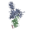
Title
Focused cryo-EM map of DDB1dB:CRBN:mezigdomide:SALL4(392-449)
Map data
Focused map, auto-sharpened with cryoSPARC
Sample
Complex: Ternary complex of DDB1dB:CRBN:mezigdomide:SALL4(392-449)
Protein or peptide: Sal-like protein 4
Protein or peptide: DNA damage-binding protein 1
Protein or peptide: Protein cereblon
Keywords
DDB1 / cereblon / mezigdomide / SALL4 / LIGASE
Function / homology
Function and homology information
POU5F1 (OCT4), SOX2, NANOG activate genes related to proliferation / Transcriptional regulation of pluripotent stem cells / negative regulation of monoatomic ion transmembrane transport / positive regulation by virus of viral protein levels in host cell / spindle assembly involved in female meiosis / embryonic limb morphogenesis / epigenetic programming in the zygotic pronuclei / ventricular septum development / inner cell mass cell proliferation / UV-damage excision repair ...POU5F1 (OCT4), SOX2, NANOG activate genes related to proliferation / Transcriptional regulation of pluripotent stem cells / negative regulation of monoatomic ion transmembrane transport / positive regulation by virus of viral protein levels in host cell / spindle assembly involved in female meiosis / embryonic limb morphogenesis / epigenetic programming in the zygotic pronuclei / ventricular septum development / inner cell mass cell proliferation / UV-damage excision repair / biological process involved in interaction with symbiont / limb development / regulation of mitotic cell cycle phase transition / WD40-repeat domain binding / Cul4A-RING E3 ubiquitin ligase complex / Cul4-RING E3 ubiquitin ligase complex / Cul4B-RING E3 ubiquitin ligase complex / ubiquitin ligase complex scaffold activity / negative regulation of reproductive process / negative regulation of developmental process / locomotory exploration behavior / somatic stem cell population maintenance / viral release from host cell / cullin family protein binding / ectopic germ cell programmed cell death / positive regulation of viral genome replication / positive regulation of Wnt signaling pathway / negative regulation of protein-containing complex assembly / heterochromatin / proteasomal protein catabolic process / positive regulation of gluconeogenesis / Regulation of PTEN gene transcription / nucleotide-excision repair / neural tube closure / sperm end piece / positive regulation of protein-containing complex assembly / regulation of circadian rhythm / Recognition of DNA damage by PCNA-containing replication complex / DNA Damage Recognition in GG-NER / Dual Incision in GG-NER / Wnt signaling pathway / Transcription-Coupled Nucleotide Excision Repair (TC-NER) / Formation of TC-NER Pre-Incision Complex / Formation of Incision Complex in GG-NER / Dual incision in TC-NER / positive regulation of protein catabolic process / Gap-filling DNA repair synthesis and ligation in TC-NER / cellular response to UV / rhythmic process / site of double-strand break / sperm principal piece / Neddylation / sperm midpiece / Potential therapeutics for SARS / ubiquitin-dependent protein catabolic process / damaged DNA binding / transmembrane transporter binding / proteasome-mediated ubiquitin-dependent protein catabolic process / DNA-binding transcription factor activity, RNA polymerase II-specific / protein-macromolecule adaptor activity / chromosome, telomeric region / protein ubiquitination / DNA repair / apoptotic process / DNA damage response / regulation of transcription by RNA polymerase II / negative regulation of apoptotic process / protein-containing complex binding / nucleolus / perinuclear region of cytoplasm / negative regulation of transcription by RNA polymerase II / positive regulation of transcription by RNA polymerase II / protein-containing complex / : / DNA binding / extracellular exosome / zinc ion binding / nucleoplasm / membrane / metal ion binding / nucleus / cytoplasm / cytosol Similarity search - Function
: / Yippee/Mis18/Cereblon / Yippee zinc-binding/DNA-binding /Mis18, centromere assembly / CULT domain / CULT domain profile. / Lon N-terminal domain profile. / Lon protease, N-terminal domain / Lon protease, N-terminal domain superfamily / ATP-dependent protease La (LON) substrate-binding domain / Found in ATP-dependent protease La (LON) ...: / Yippee/Mis18/Cereblon / Yippee zinc-binding/DNA-binding /Mis18, centromere assembly / CULT domain / CULT domain profile. / Lon N-terminal domain profile. / Lon protease, N-terminal domain / Lon protease, N-terminal domain superfamily / ATP-dependent protease La (LON) substrate-binding domain / Found in ATP-dependent protease La (LON) / : / RSE1/DDB1/CPSF1 second beta-propeller / Cleavage/polyadenylation specificity factor, A subunit, C-terminal / Cleavage/polyadenylation specificity factor, A subunit, N-terminal / : / CPSF A subunit region / RSE1/DDB1/CPSF1 first beta-propeller / PUA-like superfamily / Zinc finger, C2H2 type / zinc finger / Zinc finger C2H2 type domain profile. / Zinc finger C2H2 superfamily / Zinc finger C2H2 type domain signature. / Zinc finger C2H2-type / WD40-repeat-containing domain superfamily / WD40/YVTN repeat-like-containing domain superfamily Similarity search - Domain/homology
National Institutes of Health/National Cancer Institute (NIH/NCI)
R01CA214608
United States
Citation
Journal: Mol Cell / Year: 2025 Title: Expanding the druggable zinc-finger proteome defines properties of drug-induced degradation. Authors: Mikołaj Słabicki / Jiho Park / Radosław P Nowak / Shourya S Roy Burman / Jesse Pellman / Charles Zou / Hlib Razumkov / Jeannie Carreiro / Simran Rastogi / Anna Goldstein / Marek M Nagiec ...Authors: Mikołaj Słabicki / Jiho Park / Radosław P Nowak / Shourya S Roy Burman / Jesse Pellman / Charles Zou / Hlib Razumkov / Jeannie Carreiro / Simran Rastogi / Anna Goldstein / Marek M Nagiec / Katherine A Donovan / Jianwei Che / Moritz Hunkeler / Qixiang Geng / Chi-Lin Hsu / Megha Lakshminarayan / Chelsea Shu / Rebecca L Zon / Zuzanna Kozicka / Paul M C Park / Jonathan M Tsai / Hojong Yoon / Lyn H Jones / Adam S Sperling / Nathanael S Gray / Eric S Fischer / Benjamin L Ebert / Abstract: Glutarimide analogs, such as thalidomide, redirect the E3 ubiquitin ligase CRL4 to induce degradation of certain zinc finger (ZF) proteins. Although the core structural motif recognized by CRBN has ...Glutarimide analogs, such as thalidomide, redirect the E3 ubiquitin ligase CRL4 to induce degradation of certain zinc finger (ZF) proteins. Although the core structural motif recognized by CRBN has been characterized, it does not fully explain substrate specificity. To explore the role of residues adjacent to this core motif, we constructed a comprehensive ZF reporter library of 9,097 reporters derived from 1,655 human ZF proteins and conducted a library-on-library screen with 29 glutarimide analogs to identify compounds that collectively degrade 38 ZF reporters. Cryo-electron microscopy and crystal structures of ZFs in complex with CRBN revealed the importance of interactions beyond the core ZF degron. We used systematic mutagenesis of ZFs and CRBN to identify modes of neosubstrate recruitment requiring distinct amino acids. Finally, we found subtle chemical variations in glutarimide analogs that alter target scope and selectivity, thus providing a roadmap for their rational design.
Name: DNA damage-binding protein 1 / type: protein_or_peptide / ID: 2 Details: His6-TEV-DDB1(1-1140), with residues 396-705 replaced with a GNGNSG linker,His6-TEV-DDB1(1-1140), with residues 396-705 replaced with a GNGNSG linker Enantiomer: LEVO
Details: 50 mM HEPES/NaOH pH 7.4, 150 mM NaCl, 3 mM TCEP
Grid
Model: Quantifoil R0.6/1 / Material: GOLD / Mesh: 300 / Support film - Material: GOLD / Support film - topology: HOLEY / Support film - Film thickness: 50 / Pretreatment - Type: GLOW DISCHARGE / Pretreatment - Time: 120 sec. / Pretreatment - Atmosphere: AIR / Pretreatment - Pressure: 0.039 kPa
Vitrification
Cryogen name: ETHANE / Chamber humidity: 90 % / Chamber temperature: 283 K / Instrument: LEICA EM GP Details: UltrAuFoil R 0.6/1 300 mesh grids were glow-discharged for 2 min at 20 mA and 39 Pa, pre-incubated with 4 uL of 10 uM FLAG-IKZF1(140-196;Q146A/G151N) for 1 min, and blotted from behind for 4 ...Details: UltrAuFoil R 0.6/1 300 mesh grids were glow-discharged for 2 min at 20 mA and 39 Pa, pre-incubated with 4 uL of 10 uM FLAG-IKZF1(140-196;Q146A/G151N) for 1 min, and blotted from behind for 4 s. 4 uL of the sample was then applied to the grid. Grids were vitrified using an EM GP plunge freezer operated at 90% humidity and 10 C with 0 s pre-blot, 4 s blot, and 0 s post-blot..
Details
15 uM His6-DDB1dB:FLAG-Spy-CRBN, 150 uM mezigdomide, and 30 uM StrepII-Avi-SALL4(392-449) were mixed in dilution buffer (50 mM HEPES/NaOH pH 7.4, 150 mM NaCl, 3 mM TCEP) and incubated for 1 hr. Additional dilution buffer was added to the mixture to achieve final concentrations of 1.5 uM DDB1dB:CRBN, 15.0 uM mezigdomide, and 3.0 uM SALL4(392-449), and 4 uL of the diluted mixture were applied to the grid.
-
Electron microscopy
Microscope
TFS TALOS
Image recording
Film or detector model: GATAN K3 (6k x 4k) / Number grids imaged: 1 / Number real images: 3024 / Average exposure time: 4.8 sec. / Average electron dose: 51.7 e/Å2 Details: One movie was recorded per hole with nine holes per stage position
Electron beam
Acceleration voltage: 200 kV / Electron source: FIELD EMISSION GUN
Type of model: OTHER / Details: Ab-initio reconstruction
Final reconstruction
Applied symmetry - Point group: C1 (asymmetric) / Algorithm: FOURIER SPACE / Resolution.type: BY AUTHOR / Resolution: 3.02 Å / Resolution method: FSC 0.143 CUT-OFF / Software - Name: cryoSPARC (ver. 4.2.0) Details: Particles were re-extracted at 1.10 A/pixel, and subsequently processed by reference-based motion correction, local CTF refinement (per-particle defocus), and global CTF refinement (per- ...Details: Particles were re-extracted at 1.10 A/pixel, and subsequently processed by reference-based motion correction, local CTF refinement (per-particle defocus), and global CTF refinement (per-group beam tilt and trefoil). Homogeneous refinement of these processed particles followed by local refinement with a mask encompassing the full particle yielded a final reconstruction at 2.7 A. A local refinement using a mask encompassing the CRBN:drug:ZF portion further improved drug and ZF density. Number images used: 539282
Initial angle assignment
Type: MAXIMUM LIKELIHOOD / Software - Name: cryoSPARC (ver. 4.2.0)
Final angle assignment
Type: MAXIMUM LIKELIHOOD / Software - Name: cryoSPARC (ver. 4.2.0)
Final 3D classification
Software - Name: cryoSPARC (ver. 4.2.0) Details: Unmasked 3D classification into 20 classes with an online expectation maximization (O-EM) learning rate of 0.2 yielded 4 populated classes and 16 empty classes. Particles from 2 classes with ...Details: Unmasked 3D classification into 20 classes with an online expectation maximization (O-EM) learning rate of 0.2 yielded 4 populated classes and 16 empty classes. Particles from 2 classes with the most defined ZF density were retained for homogeneous refinement, leading to a reconstruction at 3.1 A.
chain_id: D, source_name: PDB, initial_model_type: experimental model
Details
Sharpened and unsharpened maps processed by cryoSPARC, in addition to maps post-processed with DeepEMhancer (v0.16), were used for model building. Models of DDB1dB:CRBN (PDB: 8TNQ) and SALL4(ZF3) (PDB: 8U17) were fit into the density as individual chains using ChimeraX (v1.6.1). Mezigdomide from a model of DDB1:CRBN:mezigdomide:IKZF1(ZF1-3) (PDB: 8D7Z) was added after aligning both models on CRBN, and residues outside of SALL4(ZF3) were built using Coot (v0.9.8). A ligand restraint file for mezigdomide was generated using the Grade2 (v1.6.0), and the entire model was then relaxed into the density using Rosetta (v3.13) followed by manual adjustment in Coot. The model was prepared for refinement using phenix.ready_set (v1.21-5207) and refined using phenix.real_space_refine (v1.21-5207).
Refinement
Space: REAL
+
About Yorodumi
-
News
-
Feb 9, 2022. New format data for meta-information of EMDB entries
New format data for meta-information of EMDB entries
Version 3 of the EMDB header file is now the official format.
The previous official version 1.9 will be removed from the archive.
In the structure databanks used in Yorodumi, some data are registered as the other names, "COVID-19 virus" and "2019-nCoV". Here are the details of the virus and the list of structure data.
Jan 31, 2019. EMDB accession codes are about to change! (news from PDBe EMDB page)
EMDB accession codes are about to change! (news from PDBe EMDB page)
The allocation of 4 digits for EMDB accession codes will soon come to an end. Whilst these codes will remain in use, new EMDB accession codes will include an additional digit and will expand incrementally as the available range of codes is exhausted. The current 4-digit format prefixed with “EMD-” (i.e. EMD-XXXX) will advance to a 5-digit format (i.e. EMD-XXXXX), and so on. It is currently estimated that the 4-digit codes will be depleted around Spring 2019, at which point the 5-digit format will come into force.
The EM Navigator/Yorodumi systems omit the EMD- prefix.
Related info.:Q: What is EMD? / ID/Accession-code notation in Yorodumi/EM Navigator
Yorodumi is a browser for structure data from EMDB, PDB, SASBDB, etc.
This page is also the successor to EM Navigator detail page, and also detail information page/front-end page for Omokage search.
The word "yorodu" (or yorozu) is an old Japanese word meaning "ten thousand". "mi" (miru) is to see.
Related info.:EMDB / PDB / SASBDB / Comparison of 3 databanks / Yorodumi Search / Aug 31, 2016. New EM Navigator & Yorodumi / Yorodumi Papers / Jmol/JSmol / Function and homology information / Changes in new EM Navigator and Yorodumi
 Movie
Movie Controller


 Open data
 Basic information
 Map data
 Sample
 Keywords
 Function and homology information
 Homo sapiens (human)
 Authors
 United States, 1 items
  Citation
 Structure visualization
 Downloads & links
 emd_72209.png
 http://ftp.pdbj.org/pub/emdb/structures/EMD-72209


 Z (Sec.)
 Y (Row.)
 X (Col.)


 Sample components
 Trichoplusia ni (cabbage looper)
 Processing
 Electron microscopy
 FIELD EMISSION GUN