Journal: bioRxiv / Year: 2026 Title: Toward a Random Background for Ligand Optimization. Abstract: Ligand optimization is central to drug discovery as hundreds of analogs might be designed and synthesized between an initial hit and a therapeutic candidate. The efficiency of this process is ...Ligand optimization is central to drug discovery as hundreds of analogs might be designed and synthesized between an initial hit and a therapeutic candidate. The efficiency of this process is unclear, at least partly because there is no random background for optimization against which to compare. Such a random background might emerge from synthetically accessible but otherwise systematic random small substitutions across starting ligands, measuring likelihood of achieving a substantial improvement in affinity/potency or other property by any single perturbation. Recent literature and ligand-affinity/potency databases suggest that perhaps 10% of analogs with minor modifications improve upon a parent's potency substantially (by ≥10-fold), but this number is clouded by reporting bias, intentional improvement, and inter-group reproducibility. To begin to establish a background expectation for ligand optimization, we comprehensively and systematically modified 18 lead molecules across six targets with single atom changes; 257 compounds were synthesized. Unexpectedly, 11.2% of these random small perturbation analogs improved potency by ≥10-fold over their parents. Conversely, these more potent analogs typically had worse pharmacokinetics (e.g. reduced metabolic stability, lower plasma free fraction). While it was possible to find analogs where the potency increase compensated for inferior exposure and half-life, resulting in more potent compounds in vivo, overall a frustrated landscape for ligand optimization is revealed. This study begins to establish a background expectation for ligand potency optimization and offers a simple strategy to do so. It also begins to quantify the challenges confronting the field in moving beyond in vitro potency.
Name: Signaling complex / type: complex / ID: 3 / Parent: 1 / Macromolecule list: #1-#3 Details: Heterotrimer complex of G alpha subunit o, G beta, and G gamma
Macromolecule #1: Guanine nucleotide-binding protein G(o) subunit alpha
Macromolecule
Name: Guanine nucleotide-binding protein G(o) subunit alpha / type: protein_or_peptide / ID: 1 / Number of copies: 1 / Enantiomer: LEVO EC number: Hydrolases; Acting on acid anhydrides; Acting on GTP to facilitate cellular and subcellular movement
Name: N-(5-methylnaphthalen-1-yl)pyridin-4-amine / type: ligand / ID: 6 / Number of copies: 1 / Formula: A1CIU
Molecular weight
Theoretical: 234.296 Da
-
Experimental details
-
Structure determination
Method
cryo EM
Processing
single particle reconstruction
Aggregation state
particle
-
Sample preparation
Concentration
2.6 mg/mL
Buffer
pH: 7.5 Component:
Concentration
Formula
Name
20.0 mM
C8H18N2O4S
HEPES
100.0 mM
NaCl
Sodium chloride
0.0075 % w/v
C47H88O22
Lauryl Maltose Neopentyl Glycol
0.0025 % w/v
C56H92O25
Glyco-diosgenin
0.001 % w/v
C31H50O4
Cholesteryl hemisuccinate
0.01 mM
C16H14N2
N-(5-methylnaphthalen-1-yl)pyridin-4-amine
Details: CHS was solubilized in LMNG and GDN prior to diluting into buffers.
Grid
Model: UltrAuFoil R1.2/1.3 / Material: GOLD / Mesh: 300 / Support film - Material: GOLD / Support film - topology: HOLEY / Pretreatment - Type: GLOW DISCHARGE / Pretreatment - Time: 30 sec. / Pretreatment - Atmosphere: OTHER / Details: 15 mA
Vitrification
Cryogen name: ETHANE / Chamber humidity: 100 % / Chamber temperature: 277.15 K / Instrument: FEI VITROBOT MARK IV
-
Electron microscopy
Microscope
TFS KRIOS
Image recording
Film or detector model: GATAN K3 (6k x 4k) / Number grids imaged: 1 / Number real images: 10305 / Average exposure time: 2.0 sec. / Average electron dose: 0.596 e/Å2
Electron beam
Acceleration voltage: 300 kV / Electron source: FIELD EMISSION GUN
Chain - Source name: PDB / Chain - Initial model type: experimental model
Refinement
Protocol: RIGID BODY FIT / Overall B value: 110.6
Output model
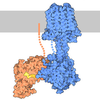
PDB-9plo: Structure of alpha2a adrenergic receptor in complex with Go heterotrimer, scFv16, and N-(5-methylnaphthalen-1-yl)pyridin-4-amine (compound 4905)
+
About Yorodumi
-
News
-
Feb 9, 2022. New format data for meta-information of EMDB entries
New format data for meta-information of EMDB entries
Version 3 of the EMDB header file is now the official format.
The previous official version 1.9 will be removed from the archive.
In the structure databanks used in Yorodumi, some data are registered as the other names, "COVID-19 virus" and "2019-nCoV". Here are the details of the virus and the list of structure data.
Jan 31, 2019. EMDB accession codes are about to change! (news from PDBe EMDB page)
EMDB accession codes are about to change! (news from PDBe EMDB page)
The allocation of 4 digits for EMDB accession codes will soon come to an end. Whilst these codes will remain in use, new EMDB accession codes will include an additional digit and will expand incrementally as the available range of codes is exhausted. The current 4-digit format prefixed with “EMD-” (i.e. EMD-XXXX) will advance to a 5-digit format (i.e. EMD-XXXXX), and so on. It is currently estimated that the 4-digit codes will be depleted around Spring 2019, at which point the 5-digit format will come into force.
The EM Navigator/Yorodumi systems omit the EMD- prefix.
Related info.:Q: What is EMD? / ID/Accession-code notation in Yorodumi/EM Navigator
Yorodumi is a browser for structure data from EMDB, PDB, SASBDB, etc.
This page is also the successor to EM Navigator detail page, and also detail information page/front-end page for Omokage search.
The word "yorodu" (or yorozu) is an old Japanese word meaning "ten thousand". "mi" (miru) is to see.
Related info.:EMDB / PDB / SASBDB / Comparison of 3 databanks / Yorodumi Search / Aug 31, 2016. New EM Navigator & Yorodumi / Yorodumi Papers / Jmol/JSmol / Function and homology information / Changes in new EM Navigator and Yorodumi
 Movie
Movie Controller

 Yorodumi
 Open data
 Basic information
 Map data
 Sample
 Keywords
 Function and homology information
 Homo sapiens (human) /
 Mus musculus (house mouse)
 Authors
 United States, 1 items
  Citation
 Structure visualization
 Downloads & links
 emd_71719.png
 https://data.pdbj.org/pub/emdb/structures/EMD-71719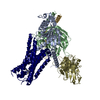





















 Z (Sec.)
 Y (Row.)
 X (Col.)


 Sample components
 Trichoplusia ni (cabbage looper)
 Spodoptera frugiperda (fall armyworm)
 Processing
 Electron microscopy
 FIELD EMISSION GUN


