 Movie
Movie Controller

[English] 日本語
 Yorodumi
- EMDB-6994: The asymmetric cryo-EM reconstruction of filamentous bacteriophage IKe -
+ Open data

- Basic information
| Entry | Database: EMDB / ID: EMD-6994 | ||||||||||||
|---|---|---|---|---|---|---|---|---|---|---|---|---|---|
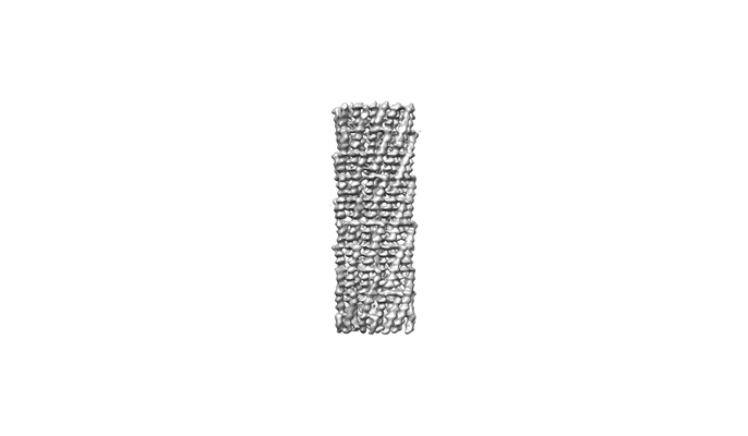
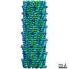
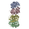
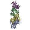
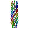
| Title | The asymmetric cryo-EM reconstruction of filamentous bacteriophage IKe | ||||||||||||
 Map data Map data | The asymmetric reconstruction of filamentous bacteriophage IKe | ||||||||||||
 Sample Sample |
| ||||||||||||
| Biological species |  Filamentous phage (virus) Filamentous phage (virus) | ||||||||||||
| Method | helical reconstruction / cryo EM / Resolution: 4.5 Å | ||||||||||||
 Authors Authors | Xu JW / Dayan N / Goldbourt A / Xiang Y | ||||||||||||
| Funding support |  China, 3 items China, 3 items
| ||||||||||||
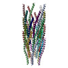
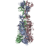
 Citation Citation | Journal: Proc Natl Acad Sci U S A / Year: 2019 Title: Cryo-electron microscopy structure of the filamentous bacteriophage IKe. Authors: Jingwei Xu / Nir Dayan / Amir Goldbourt / Ye Xiang /  Abstract: The filamentous bacteriophage IKe infects cells bearing IncN pili. We report the cryo-electron microscopy structure of the micrometer-long IKe viral particle at a resolution of 3.4 Å. The major ...The filamentous bacteriophage IKe infects cells bearing IncN pili. We report the cryo-electron microscopy structure of the micrometer-long IKe viral particle at a resolution of 3.4 Å. The major coat protein [protein 8 (p8)] consists of 47 residues that fold into a ∼68-Å-long helix. An atomic model of the coat protein was built. Five p8 helices in a horizontal layer form a pentamer, and symmetrically neighboring p8 layers form a right-handed helical cylinder having a rise per pentamer of 16.77 Å and a twist of 38.52°. The inner surface of the capsid cylinder is positively charged and has direct interactions with the encapsulated circular single-stranded DNA genome, which has an electron density consistent with an unusual left-handed helix structure. Similar to capsid structures of other filamentous viruses, strong capsid packing in the IKe particle is maintained by hydrophobic residues. Despite having a different length and large sequence differences from other filamentous phages, π-π interactions were found between Tyr9 of one p8 and Trp29 of a neighboring p8 in IKe that are similar to interactions observed in phage M13, suggesting that, despite sequence divergence, overall structural features are maintained. | ||||||||||||
| History |
|
- Structure visualization
| Movie |
 Movie viewer Movie viewer |
|---|---|
| Structure viewer | EM map: SurfViewMolmilJmol/JSmol |
| Supplemental images |
- Downloads & links
-EMDB archive
| Map data | emd_6994.map.gz | 3.6 MB | EMDB map data format | |
|---|---|---|---|---|
| Header (meta data) | emd-6994-v30.xmlemd-6994.xml | 11.8 KB 11.8 KB | Display Display | EMDB header |
| Images |  emd_6994.png emd_6994.png | 17.9 KB | ||
| Archive directory |  http://ftp.pdbj.org/pub/emdb/structures/EMD-6994ftp://ftp.pdbj.org/pub/emdb/structures/EMD-6994 http://ftp.pdbj.org/pub/emdb/structures/EMD-6994ftp://ftp.pdbj.org/pub/emdb/structures/EMD-6994 | HTTPS FTP |
-Related structure data


-Links
| EMDB pages | EMDB (EBI/PDBe) / EMDataResource |
|---|
-Map
| File | Download / File: emd_6994.map.gz / Format: CCP4 / Size: 52.7 MB / Type: IMAGE STORED AS FLOATING POINT NUMBER (4 BYTES) | ||||||||||||||||||||||||||||||||||||||||||||||||||||||||||||||||||||
|---|---|---|---|---|---|---|---|---|---|---|---|---|---|---|---|---|---|---|---|---|---|---|---|---|---|---|---|---|---|---|---|---|---|---|---|---|---|---|---|---|---|---|---|---|---|---|---|---|---|---|---|---|---|---|---|---|---|---|---|---|---|---|---|---|---|---|---|---|---|
| Annotation | The asymmetric reconstruction of filamentous bacteriophage IKe | ||||||||||||||||||||||||||||||||||||||||||||||||||||||||||||||||||||
| Projections & slices | Image control
Images are generated by Spider. | ||||||||||||||||||||||||||||||||||||||||||||||||||||||||||||||||||||
| Voxel size | X=Y=Z: 1.3 Å | ||||||||||||||||||||||||||||||||||||||||||||||||||||||||||||||||||||
| Density |
| ||||||||||||||||||||||||||||||||||||||||||||||||||||||||||||||||||||
| Symmetry | Space group: 1 | ||||||||||||||||||||||||||||||||||||||||||||||||||||||||||||||||||||
| Details | EMDB XML:
CCP4 map header:
| ||||||||||||||||||||||||||||||||||||||||||||||||||||||||||||||||||||
 Z (Sec.)
 Y (Row.)
 X (Col.)


-Supplemental data
- Sample components
-Entire : Filamentous phage
| Entire | Name: Filamentous phage (virus) |
|---|---|
| Components |
|
-Supramolecule #1: Filamentous phage
| Supramolecule | Name: Filamentous phage / type: virus / ID: 1 / Parent: 0 / Macromolecule list: all / NCBI-ID: 12420 / Sci species name: Filamentous phage / Sci species strain: IKe / Virus type: VIRION / Virus isolate: SPECIES / Virus enveloped: No / Virus empty: No |
|---|---|
| Host (natural) | Organism:  |
| Virus shell | Shell ID: 1 / Diameter: 64.0 Å |
-Macromolecule #1: The asymmetric reconstruction of filamentous bacteriophage IKe, i...
| Macromolecule | Name: The asymmetric reconstruction of filamentous bacteriophage IKe, including major coat protein shell and inner DNA core. type: other / ID: 1 / Classification: other |
|---|---|
| Source (natural) | Organism: Filamentous phage (virus) / Strain: IKe |
| Sequence | String: AEPNAATNYA TEAMDSLKTQ AIDLISQTWP VVTTVVVAGL VIRLFKKFSS KAV |
-Experimental details
-Structure determination
| Method | cryo EM |
|---|---|
 Processing Processing | helical reconstruction |
| Aggregation state | filament |
-Sample preparation
| Buffer | pH: 7.5 / Details: 50 mM Tris at pH 7.5, 10 mM MgCl2, 100 mM NaCl |
|---|---|
| Grid | Model: Quantifoil R1.2/1.3 / Material: COPPER / Mesh: 200 |
| Vitrification | Cryogen name: ETHANE / Instrument: FEI VITROBOT MARK IV |
- Electron microscopy
| Microscope | FEI TITAN KRIOS |
|---|---|
| Image recording | Film or detector model: GATAN K2 SUMMIT (4k x 4k) / Detector mode: SUPER-RESOLUTION / Average electron dose: 40.0 e/Å2 |
| Electron beam | Acceleration voltage: 300 kV / Electron source:  FIELD EMISSION GUN FIELD EMISSION GUN |
| Electron optics | Illumination mode: FLOOD BEAM / Imaging mode: BRIGHT FIELD / Cs: 2.7 mm |
| Experimental equipment |  Model: Titan Krios / Image courtesy: FEI Company |
-Image processing
| Final reconstruction | Applied symmetry - Helical parameters - Δz: 16.77 Å Applied symmetry - Helical parameters - Δ&Phi: 38.52 ° Applied symmetry - Helical parameters - Axial symmetry: C1 (asymmetric) Resolution.type: BY AUTHOR / Resolution: 4.5 Å / Resolution method: FSC 0.143 CUT-OFF / Software - Name: RELION (ver. 2.0) Details: The asymmetric reconstruction of filamentous bacteriophage IKe. The five equivalent orientation positions are generated after helical reconstruction (EMD-6993). The capsid density subtracted ...Details: The asymmetric reconstruction of filamentous bacteriophage IKe. The five equivalent orientation positions are generated after helical reconstruction (EMD-6993). The capsid density subtracted images are performed no sampling 3D classification. The best particle with high likelihood value are used for asymmetric reconstruction, which including outer helical shell and inner DNA core. Number images used: 53784 |
|---|---|
| CTF correction | Software - Name: Gctf |
| Startup model | Type of model: PDB ENTRY PDB model - PDB ID: |
| Final angle assignment | Type: NOT APPLICABLE / Software - Name: RELION (ver. 2) |
-Atomic model buiding 1
| Refinement | Space: REAL / Protocol: AB INITIO MODEL |
|---|
