 Movie
Movie Controller


+ Open data

- Basic information
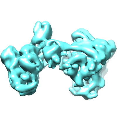
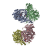
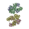
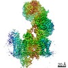
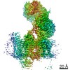
| Entry | Database: EMDB / ID: EMD-6943 | |||||||||
|---|---|---|---|---|---|---|---|---|---|---|
| Title | Saccharomyces cerevisiae Origin Recognition Complex | |||||||||
 Map data Map data | ||||||||||
 Sample Sample |
| |||||||||
| Biological species |  | |||||||||
| Method | single particle reconstruction / cryo EM / Resolution: 8.2 Å | |||||||||
 Authors Authors | Li N / Lam WH / Zhai Y / Cheng J / Cheng E / Zhao Y / Gao N / Tye BK | |||||||||
 Citation Citation | Journal: Nature / Year: 2018 Title: Structure of the origin recognition complex bound to DNA replication origin. Authors: Ningning Li / Wai Hei Lam / Yuanliang Zhai / Jiaxuan Cheng / Erchao Cheng / Yongqian Zhao / Ning Gao / Bik-Kwoon Tye /   Abstract: The six-subunit origin recognition complex (ORC) binds to DNA to mark the site for the initiation of replication in eukaryotes. Here we report a 3 Å cryo-electron microscopy structure of the ...The six-subunit origin recognition complex (ORC) binds to DNA to mark the site for the initiation of replication in eukaryotes. Here we report a 3 Å cryo-electron microscopy structure of the Saccharomyces cerevisiae ORC bound to a 72-base-pair origin DNA sequence that contains the ARS consensus sequence (ACS) and the B1 element. The ORC encircles DNA through extensive interactions with both phosphate backbone and bases, and bends DNA at the ACS and B1 sites. Specific recognition of thymine residues in the ACS is carried out by a conserved basic amino acid motif of Orc1 in the minor groove, and by a species-specific helical insertion motif of Orc4 in the major groove. Moreover, similar insertions into major and minor grooves are also embedded in the B1 site by basic patch motifs from Orc2 and Orc5, respectively, to contact bases and to bend DNA. This work pinpoints a conserved role of ORC in modulating DNA structure to facilitate origin selection and helicase loading in eukaryotes. | |||||||||
| History |
|
- Structure visualization
| Movie |
 Movie viewer Movie viewer |
|---|---|
| Structure viewer | EM map: SurfViewMolmilJmol/JSmol |
| Supplemental images |
- Downloads & links
-EMDB archive
| Map data | emd_6943.map.gz | 326.7 KB | EMDB map data format | |
|---|---|---|---|---|
| Header (meta data) | emd-6943-v30.xmlemd-6943.xml | 10 KB 10 KB | Display Display | EMDB header |
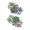
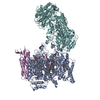
| Images |  emd_6943.png emd_6943.png | 153.4 KB | ||
| Archive directory |  http://ftp.pdbj.org/pub/emdb/structures/EMD-6943ftp://ftp.pdbj.org/pub/emdb/structures/EMD-6943 http://ftp.pdbj.org/pub/emdb/structures/EMD-6943ftp://ftp.pdbj.org/pub/emdb/structures/EMD-6943 | HTTPS FTP |
-Related structure data
-Links
| EMDB pages | EMDB (EBI/PDBe) / EMDataResource |
|---|
-Map
| File | Download / File: emd_6943.map.gz / Format: CCP4 / Size: 2.8 MB / Type: IMAGE STORED AS FLOATING POINT NUMBER (4 BYTES) | ||||||||||||||||||||||||||||||||||||||||||||||||||||||||||||
|---|---|---|---|---|---|---|---|---|---|---|---|---|---|---|---|---|---|---|---|---|---|---|---|---|---|---|---|---|---|---|---|---|---|---|---|---|---|---|---|---|---|---|---|---|---|---|---|---|---|---|---|---|---|---|---|---|---|---|---|---|---|
| Projections & slices | Image control
Images are generated by Spider. | ||||||||||||||||||||||||||||||||||||||||||||||||||||||||||||
| Voxel size | X=Y=Z: 2.64 Å | ||||||||||||||||||||||||||||||||||||||||||||||||||||||||||||
| Density |
| ||||||||||||||||||||||||||||||||||||||||||||||||||||||||||||
| Symmetry | Space group: 1 | ||||||||||||||||||||||||||||||||||||||||||||||||||||||||||||
| Details | EMDB XML:
CCP4 map header:
| ||||||||||||||||||||||||||||||||||||||||||||||||||||||||||||
 Z (Sec.)
 Y (Row.)
 X (Col.)


-Supplemental data
- Sample components
-Entire : Saccharomyces cerevisiae Origin Recognition Complex
| Entire | Name: Saccharomyces cerevisiae Origin Recognition Complex |
|---|---|
| Components |
|
-Supramolecule #1: Saccharomyces cerevisiae Origin Recognition Complex
| Supramolecule | Name: Saccharomyces cerevisiae Origin Recognition Complex / type: complex / ID: 1 / Parent: 0 / Macromolecule list: #1-#8 |
|---|---|
| Source (natural) | Organism: |
| Recombinant expression | Organism: |
-Experimental details
-Structure determination
| Method | cryo EM |
|---|---|
 Processing Processing | single particle reconstruction |
| Aggregation state | particle |
-Sample preparation
| Buffer | pH: 7.6 |
|---|---|
| Grid | Model: Quantifoil R1.2/1.3 / Material: GOLD |
| Vitrification | Cryogen name: ETHANE |
- Electron microscopy
| Microscope | FEI TITAN KRIOS |
|---|---|
| Image recording | Film or detector model: GATAN K2 SUMMIT (4k x 4k) / Detector mode: SUPER-RESOLUTION / Average electron dose: 50.0 e/Å2 |
| Electron beam | Acceleration voltage: 300 kV / Electron source:  FIELD EMISSION GUN FIELD EMISSION GUN |
| Electron optics | Illumination mode: FLOOD BEAM / Imaging mode: BRIGHT FIELD |
| Sample stage | Specimen holder model: FEI TITAN KRIOS AUTOGRID HOLDER / Cooling holder cryogen: NITROGEN |
| Experimental equipment |  Model: Titan Krios / Image courtesy: FEI Company |
-Image processing
| Final reconstruction | Applied symmetry - Point group: C1 (asymmetric) / Resolution.type: BY AUTHOR / Resolution: 8.2 Å / Resolution method: FSC 0.143 CUT-OFF / Number images used: 45000 |
|---|---|
| Initial angle assignment | Type: PROJECTION MATCHING |
| Final angle assignment | Type: PROJECTION MATCHING |
