 Movie
Movie Controller


+ Open data

- Basic information
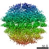
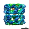
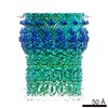
| Entry | Database: EMDB / ID: EMD-6917 | |||||||||
|---|---|---|---|---|---|---|---|---|---|---|
| Title | cryoEM structure of ETEC Pilotin-Secretin AspS-GspD complex | |||||||||
 Map data Map data | sharpening map | |||||||||
 Sample Sample |
| |||||||||
 Keywords Keywords | Pilotin / Secretin / PROTEIN TRANSPORT | |||||||||
| Function / homology |  Function and homology information Function and homology informationprotein secretion by the type II secretion system / type II protein secretion system complex / Secretion of toxins / cell outer membrane / identical protein binding Similarity search - Function | |||||||||
| Biological species |  | |||||||||
| Method | single particle reconstruction / cryo EM / Resolution: 3.2 Å | |||||||||
 Authors Authors | Yin M / Yan Z | |||||||||
 Citation Citation | Journal: Nat Microbiol / Year: 2018 Title: Structural insight into the assembly of the type II secretion system pilotin-secretin complex from enterotoxigenic Escherichia coli. Authors: Meng Yin / Zhaofeng Yan / Xueming Li /  Abstract: Secretin is a large outer-membrane channel found in secretion systems of Gram-negative bacteria, facilitating the last step for transfer of proteins into the extracellular environment. In the type II ...Secretin is a large outer-membrane channel found in secretion systems of Gram-negative bacteria, facilitating the last step for transfer of proteins into the extracellular environment. In the type II secretion system, a lipoprotein called pilotin is essential to bind and target its corresponding secretin to the outer membrane. However, there is only limited structural information available about the interaction and assembly of the pilotin-secretin complex. Here we report the first near-atomic-resolution structure of a full-length Vibrio-type pilotin-secretin (AspS-GspD) complex from enterotoxigenic Escherichia coli by cryo-electron microscopy, which reveals the detailed assembly mode of the full-length pilotin-secretin complex. The AspS subunits attach to the secretin channel surface with a 15:15 stoichiometric ratio to GspD subunits, and insert their amino terminus into the outer membrane. The AspS subunits interact with all three secondary structural elements of the S domain of GspD, including strong interaction with the carboxy-terminal α-helix and weak interactions with another two elements, an α-helix and a loop. These structural and biochemical details provide a deeper insight to pilotin-secretin interaction and their assembly mode. | |||||||||
| History |
|
- Structure visualization
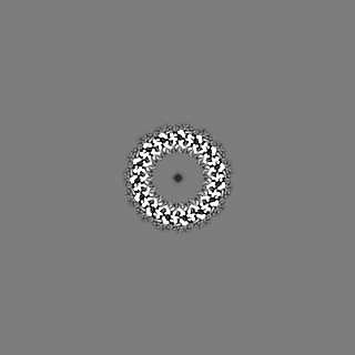
| Movie |
Movie viewer |
|---|---|
| Structure viewer | EM map: SurfViewMolmilJmol/JSmol |
| Supplemental images |

- Downloads & links
-EMDB archive
| Map data | emd_6917.map.gz | 11.6 MB | EMDB map data format | |
|---|---|---|---|---|
| Header (meta data) | emd-6917-v30.xmlemd-6917.xml | 10 KB 10 KB | Display Display | EMDB header |
| Images | 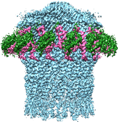 emd_6917.png emd_6917.png | 276.8 KB | ||
| Filedesc metadata | emd-6917.cif.gz | 5 KB | ||
| Archive directory |  http://ftp.pdbj.org/pub/emdb/structures/EMD-6917ftp://ftp.pdbj.org/pub/emdb/structures/EMD-6917 http://ftp.pdbj.org/pub/emdb/structures/EMD-6917ftp://ftp.pdbj.org/pub/emdb/structures/EMD-6917 | HTTPS FTP |
-Related structure data
| Related structure data |  5zdhMC  6916C M: atomic model generated by this map C: citing same article ( |
|---|---|
| Similar structure data |

-Links
| EMDB pages | EMDB (EBI/PDBe) / EMDataResource |
|---|---|
| Related items in Molecule of the Month |


-Map
| File | Download / File: emd_6917.map.gz / Format: CCP4 / Size: 125 MB / Type: IMAGE STORED AS FLOATING POINT NUMBER (4 BYTES) | ||||||||||||||||||||||||||||||||||||||||||||||||||||||||||||
|---|---|---|---|---|---|---|---|---|---|---|---|---|---|---|---|---|---|---|---|---|---|---|---|---|---|---|---|---|---|---|---|---|---|---|---|---|---|---|---|---|---|---|---|---|---|---|---|---|---|---|---|---|---|---|---|---|---|---|---|---|---|
| Annotation | sharpening map | ||||||||||||||||||||||||||||||||||||||||||||||||||||||||||||
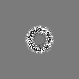
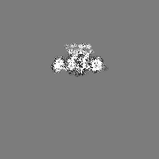
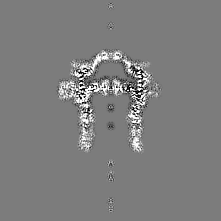
| Projections & slices | Image control
Images are generated by Spider. | ||||||||||||||||||||||||||||||||||||||||||||||||||||||||||||
| Voxel size | X=Y=Z: 1.32 Å | ||||||||||||||||||||||||||||||||||||||||||||||||||||||||||||
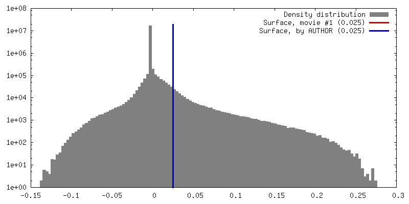
| Density |
| ||||||||||||||||||||||||||||||||||||||||||||||||||||||||||||
| Symmetry | Space group: 1 | ||||||||||||||||||||||||||||||||||||||||||||||||||||||||||||
| Details | EMDB XML:
CCP4 map header:
| ||||||||||||||||||||||||||||||||||||||||||||||||||||||||||||
 Z (Sec.)
 Y (Row.)
 X (Col.)


-Supplemental data
- Sample components
-Entire : Pilotin-Secretin Complex Asps-GspD
| Entire | Name: Pilotin-Secretin Complex Asps-GspD |
|---|---|
| Components |
|
-Supramolecule #1: Pilotin-Secretin Complex Asps-GspD
| Supramolecule | Name: Pilotin-Secretin Complex Asps-GspD / type: complex / ID: 1 / Parent: 0 / Macromolecule list: all |
|---|---|
| Source (natural) | Organism: |
-Macromolecule #1: Type II secretion system protein D
| Macromolecule | Name: Type II secretion system protein D / type: protein_or_peptide / ID: 1 / Number of copies: 15 / Enantiomer: LEVO |
|---|---|
| Source (natural) | Organism: Strain: H10407 / ETEC |
| Molecular weight | Theoretical: 69.70082 KDa |
| Recombinant expression | Organism: |
| Sequence | String: EEATFTANFK DTDLKSFIET VGANLNKTII MGPGVQGKVS IRTMTPLNER QYYQLFLNLL EAQGYAVVPM ENDVLKVVKS SAAKVEPLP LVGEGSDNYA GDEMVTKVVP VRNVSVRELA PILRQMIDSA GSGNVVNYDP SNVIMLTGRA SVVERLTEVI Q RVDHAGNR ...String: EEATFTANFK DTDLKSFIET VGANLNKTII MGPGVQGKVS IRTMTPLNER QYYQLFLNLL EAQGYAVVPM ENDVLKVVKS SAAKVEPLP LVGEGSDNYA GDEMVTKVVP VRNVSVRELA PILRQMIDSA GSGNVVNYDP SNVIMLTGRA SVVERLTEVI Q RVDHAGNR TEEVIPLDNA SASEIARVLE SLTKNSGENQ PATLKSQIVA DERTNSVIVS GDPATRDKMR RLIRRLDSEM ER SGNSQVF YLKYSKAEDL VDVLKQVSGT LTAAKEEAEG TVGSGREIVS IAASKHSNAL IVTAPQDIMQ SLQSVIEQLD IRR AQVHVE ALIVEVAEGS NINFGVQWAS KDAGLMQFAN GTQIPIGTLG AAISQAKPQK GSTVISENGA TTINPDTNGD LSTL AQLLS GFSGTAVGVV KGDWMALVQA VKNDSSSNVL STPSITTLDN QEAFFMVGQD VPVLTGSTVG SNNSNPFNTV ERKKV GIML KVTPQINEGN AVQMVIEQEV SKVEGQTSLD VVFGERKLKT TVLANDGELI VLGGLMDDQA GESVAKVPLL GDIPLI GNL FKSTADKKEK RNLMVFIRPT ILRDGMAADG VSQRKYNYMR AEQIYRDEQG LSLMPHTAQP VLPAQNQALP PEVRAFL NA GRTR UniProtKB: Secretin GspD 2 |
-Macromolecule #2: Type II secretion system lipoprotein
| Macromolecule | Name: Type II secretion system lipoprotein / type: protein_or_peptide / ID: 2 / Number of copies: 15 / Enantiomer: LEVO |
|---|---|
| Source (natural) | Organism: Strain: H10407 / ETEC |
| Molecular weight | Theoretical: 12.18578 KDa |
| Recombinant expression | Organism: |
| Sequence | String: CASHNENASL LAKKQAQNIS QNLPIKSAGY TLVLAQSSGT TVKMTIISEA GTQTTQTPDA FLTSYQRQMC ADPTVKLMLT EGINYSITI NDTRTGNQYQ RKLDRTTCGI VKA UniProtKB: Pilotin AspS 2 |
-Experimental details
-Structure determination
| Method | cryo EM |
|---|---|
 Processing Processing | single particle reconstruction |
| Aggregation state | particle |
-Sample preparation
| Buffer | pH: 8 |
|---|---|
| Vitrification | Cryogen name: ETHANE |
- Electron microscopy
| Microscope | FEI TITAN KRIOS |
|---|---|
| Image recording | Film or detector model: GATAN K2 SUMMIT (4k x 4k) / Average electron dose: 50.0 e/Å2 |
| Electron beam | Acceleration voltage: 300 kV / Electron source:  FIELD EMISSION GUN FIELD EMISSION GUN |
| Electron optics | Illumination mode: OTHER / Imaging mode: BRIGHT FIELD |
| Experimental equipment |  Model: Titan Krios / Image courtesy: FEI Company |
-Image processing
| Startup model | Type of model: OTHER |
|---|---|
| Final reconstruction | Resolution.type: BY AUTHOR / Resolution: 3.2 Å / Resolution method: FSC 0.143 CUT-OFF / Number images used: 37928 |
| Initial angle assignment | Type: OTHER |
| Final angle assignment | Type: OTHER |
