 Movie
Movie Controller


+ Open data

- Basic information
| Entry | Database: EMDB / ID: EMD-6916 | |||||||||
|---|---|---|---|---|---|---|---|---|---|---|
| Title | CryoEM structure of ETEC Secretin GspD | |||||||||
 Map data Map data | sharpening map | |||||||||
 Sample Sample |
| |||||||||
| Biological species |  | |||||||||
| Method | single particle reconstruction / cryo EM / Resolution: 3.91 Å | |||||||||
 Authors Authors | Yin M / Yan Z / Li X | |||||||||
 Citation Citation | Journal: Nat Microbiol / Year: 2018 Title: Structural insight into the assembly of the type II secretion system pilotin-secretin complex from enterotoxigenic Escherichia coli. Authors: Meng Yin / Zhaofeng Yan / Xueming Li /  Abstract: Secretin is a large outer-membrane channel found in secretion systems of Gram-negative bacteria, facilitating the last step for transfer of proteins into the extracellular environment. In the type II ...Secretin is a large outer-membrane channel found in secretion systems of Gram-negative bacteria, facilitating the last step for transfer of proteins into the extracellular environment. In the type II secretion system, a lipoprotein called pilotin is essential to bind and target its corresponding secretin to the outer membrane. However, there is only limited structural information available about the interaction and assembly of the pilotin-secretin complex. Here we report the first near-atomic-resolution structure of a full-length Vibrio-type pilotin-secretin (AspS-GspD) complex from enterotoxigenic Escherichia coli by cryo-electron microscopy, which reveals the detailed assembly mode of the full-length pilotin-secretin complex. The AspS subunits attach to the secretin channel surface with a 15:15 stoichiometric ratio to GspD subunits, and insert their amino terminus into the outer membrane. The AspS subunits interact with all three secondary structural elements of the S domain of GspD, including strong interaction with the carboxy-terminal α-helix and weak interactions with another two elements, an α-helix and a loop. These structural and biochemical details provide a deeper insight to pilotin-secretin interaction and their assembly mode. | |||||||||
| History |
|
- Structure visualization
| Movie |
 Movie viewer Movie viewer |
|---|---|
| Structure viewer | EM map: SurfViewMolmilJmol/JSmol |
| Supplemental images |

- Downloads & links
-EMDB archive
| Map data | emd_6916.map.gz | 8.7 MB | EMDB map data format | |
|---|---|---|---|---|
| Header (meta data) | emd-6916-v30.xmlemd-6916.xml | 9 KB 9 KB | Display Display | EMDB header |
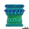
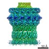
| Images | 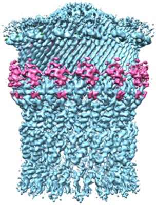 emd_6916.png emd_6916.png | 245.7 KB | ||
| Archive directory |  http://ftp.pdbj.org/pub/emdb/structures/EMD-6916ftp://ftp.pdbj.org/pub/emdb/structures/EMD-6916 http://ftp.pdbj.org/pub/emdb/structures/EMD-6916ftp://ftp.pdbj.org/pub/emdb/structures/EMD-6916 | HTTPS FTP |
-Related structure data


-Links
| EMDB pages | EMDB (EBI/PDBe) / EMDataResource |
|---|
-Map
| File | Download / File: emd_6916.map.gz / Format: CCP4 / Size: 125 MB / Type: IMAGE STORED AS FLOATING POINT NUMBER (4 BYTES) | ||||||||||||||||||||||||||||||||||||||||||||||||||||||||||||
|---|---|---|---|---|---|---|---|---|---|---|---|---|---|---|---|---|---|---|---|---|---|---|---|---|---|---|---|---|---|---|---|---|---|---|---|---|---|---|---|---|---|---|---|---|---|---|---|---|---|---|---|---|---|---|---|---|---|---|---|---|---|
| Annotation | sharpening map | ||||||||||||||||||||||||||||||||||||||||||||||||||||||||||||
| Projections & slices | Image control
Images are generated by Spider. | ||||||||||||||||||||||||||||||||||||||||||||||||||||||||||||
| Voxel size | X=Y=Z: 1.32 Å | ||||||||||||||||||||||||||||||||||||||||||||||||||||||||||||
| Density |
| ||||||||||||||||||||||||||||||||||||||||||||||||||||||||||||
| Symmetry | Space group: 1 | ||||||||||||||||||||||||||||||||||||||||||||||||||||||||||||
| Details | EMDB XML:
CCP4 map header:
| ||||||||||||||||||||||||||||||||||||||||||||||||||||||||||||
 Z (Sec.)
 Y (Row.)
 X (Col.)


-Supplemental data
- Sample components
-Entire : GspD
| Entire | Name: GspD |
|---|---|
| Components |
|
-Supramolecule #1: GspD
| Supramolecule | Name: GspD / type: complex / ID: 1 / Parent: 0 / Macromolecule list: all / Details: outer membrane channel Secretin |
|---|---|
| Source (natural) | Organism: |
| Recombinant expression | Organism: |
-Macromolecule #1: GspD
| Macromolecule | Name: GspD / type: protein_or_peptide / ID: 1 / Enantiomer: DEXTRO |
|---|---|
| Source (natural) | Organism: |
| Recombinant expression | Organism: |
| Sequence | String: EEATFTANFK DTDLKSFIET VGANLNKTI IMGPGVQGKV SIRTMTPLNE RQYYQLFLNL LEAQGYAVVP MENDVLKVVK S SAAKVEPL PLVGEGSDNY AGDEMVTKVV PVRNVSVREL APILRQMIDS AGSGNVVNYD PS NVIMLTG RASVVERLTE VIQRVDHAGN ...String: EEATFTANFK DTDLKSFIET VGANLNKTI IMGPGVQGKV SIRTMTPLNE RQYYQLFLNL LEAQGYAVVP MENDVLKVVK S SAAKVEPL PLVGEGSDNY AGDEMVTKVV PVRNVSVREL APILRQMIDS AGSGNVVNYD PS NVIMLTG RASVVERLTE VIQRVDHAGN RTEEVIPLDN ASASEIARVL ESLTKNSGEN QPA TLKSQI VADERTNSVI VSGDPATRDK MRRLIRRLDS EMERSGNSQV FYLKYSKAED LVDV LKQVS GTLTAAKEEA EGTVGSGREI VSIAASKHSN ALIVTAPQDI MQSLQSVIEQ LDIRR AQVH VEALIVEVAE GSNINFGVQW ASKDAGLMQF ANGTQIPIGT LGAAISQAKP QKGSTV ISE NGATTINPDT NGDLSTLAQL LSGFSGTAVG VVKGDWMALV QAVKNDSSSN VLSTPSI TT LDNQEAFFMV GQDVPVLTGS TVGSNNSNPF NTVERKKVGI MLKVTPQINE GNAVQMVI E QEVSKVEGQT SLDVVFGERK LKTTVLANDG ELIVLGGLMD DQAGESVAKV PLLGDIPLI GNLFKSTADK KEKRNLMVFI RPTILRDGMA ADGVSQRKYN YMRAEQIYRD EQGLSLMPHT AQPVLPAQN QALPPEVRAF LNAGRTR |
-Experimental details
-Structure determination
| Method | cryo EM |
|---|---|
 Processing Processing | single particle reconstruction |
| Aggregation state | particle |
-Sample preparation
| Buffer | pH: 8 |
|---|---|
| Vitrification | Cryogen name: ETHANE |
- Electron microscopy
| Microscope | FEI TITAN KRIOS |
|---|---|
| Image recording | Film or detector model: GATAN K2 SUMMIT (4k x 4k) / Average electron dose: 50.0 e/Å2 |
| Electron beam | Acceleration voltage: 300 kV / Electron source:  FIELD EMISSION GUN FIELD EMISSION GUN |
| Electron optics | Illumination mode: OTHER / Imaging mode: BRIGHT FIELD |
| Experimental equipment |  Model: Titan Krios / Image courtesy: FEI Company |
-Image processing
| CTF correction | Software - Name: CTFFIND (ver. 1.4) |
|---|---|
| Final reconstruction | Resolution.type: BY AUTHOR / Resolution: 3.91 Å / Resolution method: FSC 0.143 CUT-OFF / Software - Name: RELION (ver. 1.4) / Number images used: 17528 |
| Initial angle assignment | Type: OTHER |
| Final angle assignment | Type: OTHER |
