 Movie
Movie Controller

[English] 日本語
 Yorodumi
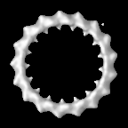
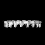
- EMDB-6912: Subtomogram average of the budding yeast DASH outer-kinetochore ring. -
+ Open data

- Basic information
| Entry | Database: EMDB / ID: EMD-6912 | |||||||||
|---|---|---|---|---|---|---|---|---|---|---|
| Title | Subtomogram average of the budding yeast DASH outer-kinetochore ring. | |||||||||
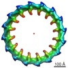
 Map data Map data | Subtomogram average of reconstituted DASH outer-kinetochore rings around microtubules. | |||||||||
 Sample Sample |
| |||||||||
| Biological species |  | |||||||||
| Method | electron tomography / cryo EM / Resolution: 32.0 Å | |||||||||
 Authors Authors | Ng C / Deng L / Chen C / Lim H / Shi J / Surana U / Gan L | |||||||||
 Citation Citation | Journal: J Cell Biol / Year: 2019 Title: Electron cryotomography analysis of Dam1C/DASH at the kinetochore-spindle interface in situ. Authors: Cai Tong Ng / Li Deng / Chen Chen / Hong Hwa Lim / Jian Shi / Uttam Surana / Lu Gan /  Abstract: In dividing cells, depolymerizing spindle microtubules move chromosomes by pulling at their kinetochores. While kinetochore subcomplexes have been studied extensively in vitro, little is known about ...In dividing cells, depolymerizing spindle microtubules move chromosomes by pulling at their kinetochores. While kinetochore subcomplexes have been studied extensively in vitro, little is known about their in vivo structure and interactions with microtubules or their response to spindle damage. Here we combine electron cryotomography of serial cryosections with genetic and pharmacological perturbation to study the yeast chromosome segregation machinery in vivo. Each kinetochore microtubule has one (rarely, two) Dam1C/DASH outer kinetochore assemblies. Dam1C/DASH contacts the microtubule walls and does so with its flexible "bridges"; there are no contacts with the protofilaments' curved tips. In metaphase, ∼40% of the Dam1C/DASH assemblies are complete rings; the rest are partial rings. Ring completeness and binding position along the microtubule are sensitive to kinetochore attachment and tension, respectively. Our study and those of others support a model in which each kinetochore must undergo cycles of conformational change to couple microtubule depolymerization to chromosome movement. | |||||||||
| History |
|
- Structure visualization
| Movie |
 Movie viewer Movie viewer |
|---|---|
| Structure viewer | EM map: SurfViewMolmilJmol/JSmol |
| Supplemental images |

- Downloads & links
-EMDB archive
| Map data | emd_6912.map.gz | 1.8 MB | EMDB map data format | |
|---|---|---|---|---|
| Header (meta data) | emd-6912-v30.xmlemd-6912.xml | 10.8 KB 10.8 KB | Display Display | EMDB header |
| Images | 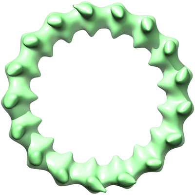 emd_6912.png emd_6912.png | 87.6 KB | ||
| Archive directory |  http://ftp.pdbj.org/pub/emdb/structures/EMD-6912ftp://ftp.pdbj.org/pub/emdb/structures/EMD-6912 http://ftp.pdbj.org/pub/emdb/structures/EMD-6912ftp://ftp.pdbj.org/pub/emdb/structures/EMD-6912 | HTTPS FTP |
-Related structure data
| Related structure data |  6914C C: citing same article ( |
|---|---|
| Similar structure data | |
| EM raw data | EMPIAR-10159 (Title: A multi-scale model of the yeast chromosome-segregation system Data size: 95.4 Data #1: Tilt series, tomograms, and models of cryosections of mitotic budding yeast and reconstituted DASH rings around microtubules [tilt series]) |
-Links
| EMDB pages | EMDB (EBI/PDBe) / EMDataResource |
|---|
-Map
| File | Download / File: emd_6912.map.gz / Format: CCP4 / Size: 11.4 MB / Type: IMAGE STORED AS FLOATING POINT NUMBER (4 BYTES) | ||||||||||||||||||||||||||||||||||||||||||||||||||||||||||||||||||||
|---|---|---|---|---|---|---|---|---|---|---|---|---|---|---|---|---|---|---|---|---|---|---|---|---|---|---|---|---|---|---|---|---|---|---|---|---|---|---|---|---|---|---|---|---|---|---|---|---|---|---|---|---|---|---|---|---|---|---|---|---|---|---|---|---|---|---|---|---|---|
| Annotation | Subtomogram average of reconstituted DASH outer-kinetochore rings around microtubules. | ||||||||||||||||||||||||||||||||||||||||||||||||||||||||||||||||||||
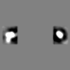
| Projections & slices | Image control
Images are generated by Spider. | ||||||||||||||||||||||||||||||||||||||||||||||||||||||||||||||||||||
| Voxel size | X=Y=Z: 4.61 Å | ||||||||||||||||||||||||||||||||||||||||||||||||||||||||||||||||||||
| Density |
| ||||||||||||||||||||||||||||||||||||||||||||||||||||||||||||||||||||
| Symmetry | Space group: 1 | ||||||||||||||||||||||||||||||||||||||||||||||||||||||||||||||||||||
| Details | EMDB XML:
CCP4 map header:
| ||||||||||||||||||||||||||||||||||||||||||||||||||||||||||||||||||||
 Z (Sec.)
 Y (Row.)
 X (Col.)


-Supplemental data
- Sample components
-Entire : Budding yeast DASH outer-kinetochore ring complex
| Entire | Name: Budding yeast DASH outer-kinetochore ring complex |
|---|---|
| Components |
|
-Supramolecule #1: Budding yeast DASH outer-kinetochore ring complex
| Supramolecule | Name: Budding yeast DASH outer-kinetochore ring complex / type: complex / ID: 1 / Parent: 0 |
|---|---|
| Source (natural) | Organism: |
| Molecular weight | Theoretical: 3.4 MDa |
-Experimental details
-Structure determination
| Method | cryo EM |
|---|---|
 Processing Processing | electron tomography |
| Aggregation state | particle |
-Sample preparation
| Concentration | 1 mg/mL |
|---|---|
| Buffer | pH: 6.8 Details: 20 mM sodium phosphate pH 6.8, 150 mM NaCl, 1 mM EDTA |
| Grid | Model: Quantifoil R2/2 / Material: COPPER / Mesh: 200 / Support film - Material: CARBON / Support film - topology: HOLEY |
| Vitrification | Cryogen name: ETHANE |
| Details | DASH complexes mixed with Taxol-stabilized microtubules |
| Sectioning | Other: NO SECTIONING |
| Fiducial marker | Manufacturer: BBI / Diameter: 10 nm |
- Electron microscopy
| Microscope | FEI TITAN KRIOS |
|---|---|
| Image recording | Film or detector model: FEI FALCON II (4k x 4k) / Detector mode: INTEGRATING / Digitization - Dimensions - Width: 4096 pixel / Digitization - Dimensions - Height: 4096 pixel / Digitization - Sampling interval: 14.0 µm / Number grids imaged: 1 / Number real images: 67 / Average electron dose: 1.5 e/Å2 |
| Electron beam | Acceleration voltage: 300 kV / Electron source:  FIELD EMISSION GUN FIELD EMISSION GUN |
| Electron optics | Calibrated magnification: 30400 / Illumination mode: FLOOD BEAM / Imaging mode: BRIGHT FIELD / Nominal defocus max: 14.0 µm / Nominal defocus min: 8.0 µm / Nominal magnification: 18000 |
| Sample stage | Specimen holder model: FEI TITAN KRIOS AUTOGRID HOLDER / Cooling holder cryogen: NITROGEN |
| Experimental equipment |  Model: Titan Krios / Image courtesy: FEI Company |
-Image processing
| Final reconstruction | Algorithm: BACK PROJECTION / Resolution.type: BY AUTHOR / Resolution: 32.0 Å / Resolution method: FSC 0.143 CUT-OFF Software:
Number images used: 66 | ||||||||
|---|---|---|---|---|---|---|---|---|---|
| CTF correction | Software - Name: eTomo (ver. 4.9) / Details: CTF compensation done in Etomo |
