 Movie
Movie Controller


+ Open data

- Basic information
| Entry | Database: EMDB / ID: EMD-5254 | |||||||||
|---|---|---|---|---|---|---|---|---|---|---|
| Title | WT Dam1 complex assembled into a ring around a microtubule | |||||||||
 Map data Map data | 16 WT Dam1 complexes assembled into a ring around a microtubule | |||||||||
 Sample Sample |
| |||||||||
 Keywords Keywords | Kinetochore / MAP | |||||||||
| Biological species |  | |||||||||
| Method | single particle reconstruction / cryo EM / Resolution: 35.0 Å | |||||||||
 Authors Authors | Ramey V / Wang HW / Nogales E | |||||||||
 Citation Citation | Journal: Mol Biol Cell / Year: 2011 Title: The Dam1 ring binds to the E-hook of tubulin and diffuses along the microtubule. Authors: Vincent H Ramey / Hong-Wei Wang / Yuko Nakajima / Amanda Wong / Jian Liu / David Drubin / Georjana Barnes / Eva Nogales /  Abstract: There has been much effort in recent years aimed at understanding the molecular mechanism by which the Dam1 kinetochore complex is able to couple microtubule depolymerization to poleward movement. ...There has been much effort in recent years aimed at understanding the molecular mechanism by which the Dam1 kinetochore complex is able to couple microtubule depolymerization to poleward movement. Both a biased diffusion and a forced walk model have been proposed, and several key functional aspects of Dam1-microtubule binding are disputed. Here, we investigate the elements involved in tubulin-Dam1 complex interactions and directly visualize Dam1 rings on microtubules in order to infer their dynamic behavior on the microtubule lattice and its likely relevance at the kinetochore. We find that the Dam1 complex has a preference for native tubulin over tubulin that is lacking its acidic C-terminal tail. Statistical mechanical analysis of images of Dam1 rings on microtubules, applied to both the distance between rings and the tilt angle of the rings with respect to the microtubule axis, supports a diffusive ring model. We also present a cryo-EM reconstruction of the Dam1 ring, likely the relevant assembly form of the complex for energy coupling during microtubule depolymerization in budding yeast. The present studies constitute a significant step forward by linking structural and biochemical observations toward a comprehensive understanding of the Dam1 complex. | |||||||||
| History |
|
- Structure visualization
| Movie |
 Movie viewer Movie viewer |
|---|---|
| Structure viewer | EM map: SurfViewMolmilJmol/JSmol |
| Supplemental images |

- Downloads & links
-EMDB archive
| Map data | emd_5254.map.gz | 27.1 MB | EMDB map data format | |
|---|---|---|---|---|
| Header (meta data) | emd-5254-v30.xmlemd-5254.xml | 9.8 KB 9.8 KB | Display Display | EMDB header |
| Images | 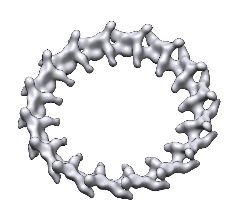 emd_5254_1.jpg emd_5254_1.jpg | 64.6 KB | ||
| Archive directory |  http://ftp.pdbj.org/pub/emdb/structures/EMD-5254ftp://ftp.pdbj.org/pub/emdb/structures/EMD-5254 http://ftp.pdbj.org/pub/emdb/structures/EMD-5254ftp://ftp.pdbj.org/pub/emdb/structures/EMD-5254 | HTTPS FTP |
-Related structure data
| Similar structure data |
|---|

-Links
| EMDB pages | EMDB (EBI/PDBe) / EMDataResource |
|---|
-Map
| File | Download / File: emd_5254.map.gz / Format: CCP4 / Size: 29.8 MB / Type: IMAGE STORED AS FLOATING POINT NUMBER (4 BYTES) | ||||||||||||||||||||||||||||||||||||||||||||||||||||||||||||||||||||
|---|---|---|---|---|---|---|---|---|---|---|---|---|---|---|---|---|---|---|---|---|---|---|---|---|---|---|---|---|---|---|---|---|---|---|---|---|---|---|---|---|---|---|---|---|---|---|---|---|---|---|---|---|---|---|---|---|---|---|---|---|---|---|---|---|---|---|---|---|---|
| Annotation | 16 WT Dam1 complexes assembled into a ring around a microtubule | ||||||||||||||||||||||||||||||||||||||||||||||||||||||||||||||||||||
| Projections & slices | Image control
Images are generated by Spider. | ||||||||||||||||||||||||||||||||||||||||||||||||||||||||||||||||||||
| Voxel size | X=Y=Z: 4.6 Å | ||||||||||||||||||||||||||||||||||||||||||||||||||||||||||||||||||||
| Density |
| ||||||||||||||||||||||||||||||||||||||||||||||||||||||||||||||||||||
| Symmetry | Space group: 1 | ||||||||||||||||||||||||||||||||||||||||||||||||||||||||||||||||||||
| Details | EMDB XML:
CCP4 map header:
| ||||||||||||||||||||||||||||||||||||||||||||||||||||||||||||||||||||
 Z (Sec.)
 Y (Row.)
 X (Col.)


-Supplemental data
- Sample components
-Entire : WT Dam1 ring
| Entire | Name: WT Dam1 ring |
|---|---|
| Components |
|
-Supramolecule #1000: WT Dam1 ring
| Supramolecule | Name: WT Dam1 ring / type: sample / ID: 1000 / Oligomeric state: 16 Dam1 complexes / Number unique components: 1 |
|---|---|
| Molecular weight | Theoretical: 210 KDa |
-Macromolecule #1: Dam1 complex
| Macromolecule | Name: Dam1 complex / type: protein_or_peptide / ID: 1 / Name.synonym: Dam1 complex / Number of copies: 16 / Oligomeric state: Ring / Recombinant expression: Yes |
|---|---|
| Source (natural) | Organism: |
| Molecular weight | Theoretical: 210 KDa |
| Recombinant expression | Organism:  |
-Experimental details
-Structure determination
| Method | cryo EM |
|---|---|
 Processing Processing | single particle reconstruction |
| Aggregation state | particle |
-Sample preparation
| Concentration | 0.01 mg/mL |
|---|---|
| Buffer | pH: 6.8 / Details: 80mM PIPES, 1mM EGTA, 1mM MgCl2 |
| Grid | Details: 400 mesh C-flat 1.2um holes |
| Vitrification | Cryogen name: ETHANE / Chamber humidity: 100 % / Chamber temperature: 77 K / Instrument: OTHER Details: Vitrification instrument: FEI vitrobot. Waited for sample to pass through open holes before blotting Method: Blot for 2 seconds before plunging |
- Electron microscopy
| Microscope | FEI TECNAI F20 |
|---|---|
| Temperature | Average: 91 K |
| Alignment procedure | Legacy - Astigmatism: objective lens astigmatism was corrected at 100,000 times magnification |
| Date | Mar 31, 2009 |
| Image recording | Category: CCD / Film or detector model: GATAN ULTRASCAN 4000 (4k x 4k) / Digitization - Sampling interval: 15 µm / Number real images: 50 / Average electron dose: 15 e/Å2 / Bits/pixel: 16 |
| Electron beam | Acceleration voltage: 120 kV / Electron source:  FIELD EMISSION GUN FIELD EMISSION GUN |
| Electron optics | Illumination mode: FLOOD BEAM / Imaging mode: BRIGHT FIELD / Cs: 2.6 mm / Nominal defocus max: 2.8 µm / Nominal defocus min: 1.17 µm / Nominal magnification: 50000 |
| Sample stage | Specimen holder: Single tilt cryo / Specimen holder model: GATAN LIQUID NITROGEN |
| Experimental equipment |  Model: Tecnai F20 / Image courtesy: FEI Company |
-Image processing
| CTF correction | Details: per micrograph |
|---|---|
| Final reconstruction | Algorithm: OTHER / Resolution.type: BY AUTHOR / Resolution: 35.0 Å / Resolution method: FSC 0.5 CUT-OFF / Software - Name: SPIDER Details: 16 fold rotational symmetry applied to reconstruction Number images used: 236 |
